Case Series
Herpes Zoster Erroneously Diagnosed as Renal Colic: A Report of 3 Cases
Ahmed Assem1, Ashraf A Mosharafa1* and Rehab Sohi2
1Departments of Urology, Cairo University, Egypt
2Departments of Dermatology, Cairo University, Egypt
*Corresponding author: Ashraf A Mosharafa, Department of Urology, Cairo University, 34 El-Nadi Street, Maadi, Cairo, 11431, Egypt
Published: 11 Oct, 2018
Cite this article as: Assem A, Mosharafa AA, Sohi R.
Herpes Zoster Erroneously Diagnosed
as Renal Colic: A Report of 3 Cases.
Ann Clin Case Rep. 2018; 3: 1548.
Abstract
Patients with Herpes Zoster presenting in the initial disease period (before the appearance of herpetic eruption) are frequently misdiagnosed. We report on three cases of Herpes Zoster initially misdiagnosed as renal colic. We describe the clinical, laboratory and imaging findings and suggest measures that could help avoid an erroneous diagnosis.
Introduction
Patients with Herpes Zoster presenting in the initial disease period (before the appearance of herpetic eruption) frequently receive an erroneous diagnosis such as renal colic, acute pancreatitis, and biliary colic [1,2]. We present our experience with three cases initially diagnosed as renal colic, and subsequently identified as cases of Herpes Zoster. A description of the clinical and investigational findings is followed by a discussion of the pitfalls leading to an erroneous diagnosis and suggestions to avoid them.
Observations
Case 1
A 37- year-old lady presented to the emergency department of our tertiary care hospital with
right loin pain of two days duration, described as dull or pricking, radiating to the spine, not related to
meals, position or exertion. A provisional diagnosis of renal colic was made and the patient received
analgesia and was referred to the Urology clinic. The patient had no lower urinary tract symptoms.
She was under treatment for infertility but had no other medical problems. On examination, she was
overweight with a lax non-tender abdomen. A urine analysis was within normal and an abdominopelvic
ultrasound revealed mild hepatomegaly. Further imaging was requested and the patient was
given a follow up appointment. Four days later, the patient presented back to the urology clinic with
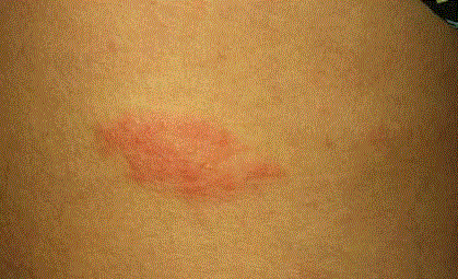
a rash that had appeared on the 5th day of pain (Figure 1), consisting of an elevated erythematous
base with multiple tiny vesicles in the right flank (T10 dermatome). The patient was referred to the
Dermatology clinic where she was started on antiviral and NSAID medications.
Case 2
A 52-year-old lady presented to the urology clinic with left loin pain, shooting forward to the
iliac region, pain score 5/10. The patient had visited the ER of her primary health facility twice
during the previous four days because of the same complaint. Pain was associated with mild urinary
frequency and dysuria. The patient was receiving treatment for a number of medical conditions
including diabetes mellitus, dyslipidemia, rheumatoid arthritis and multinodular goiter. She had
undergone cholecystectomy 22 years earlier. Her urine analysis revealed WBCs 20-30/HPF, RBCs
3-5/HPF and proteinuria. Urine analysis was negative for nitrites and bacteria. Complete blood
count revealed a total leucocytic count of 6,000/μL; Neutrophils 45%; C-reactive protein 1.2 mg/dL
(normally 0-0.5) and Erythrocyte Sedimentation Rate first hour of 74 (normally 0-20). A provisional
diagnosis of cystitis and renal pain was withheld. Urine culture and Ultrasound were requested.
On the follow up visit, her urine culture was negative; ultrasound did not reveal any urologic
abnormality. Herpes Zoster was suspected based on the appearance of a rash and the patient was
referred for further evaluation and treatment by infectious diseases clinic.
Case 3
A 51-year-old male patient presented to the urology clinic with severe right loin pain for three
days, burning in nature, associated with intermediate grade fever. The patient was a known stone former/stone passer for the previous four years. His medical history
is also significant for hypertension and history of knee replacement
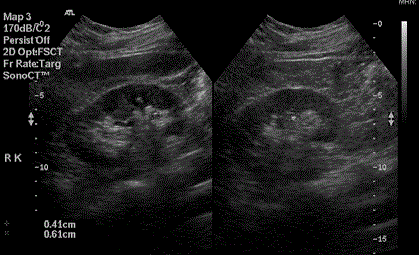
surgery. A prior Ultrasound (done 3 months before presentation)
had suggested 3 mm to 5 mm bilateral stones and a repeat ultrasound
suggested two small (4 mm and 6 mm) stones in the lower and mid
zones of the right kidney (Figure 2). The patient was admitted to the
hospital for control of pain and fever. His serum creatinine was stable
at 1.3 mg/dL, serum uric acid was 5.8 mg/dL; urine analysis showed
a few urate crystals and urine C & S was negative. A CT urinary tract
was requested and revealed no significant renal or ureteric stones
and no hydronephrosis. A rash (erythema and vesicles along the
T11 abdominal dermatome) appeared on the 5th days; Dermatology
and infectious disease consultations confirmed Herpes Zoster and
treatment was started with Voltrex 500 mg 2 tabs tds, Neurontin 300
mg PO tds with tramal and NSAID.
Figure 1
Discussion
Pain associated with Herpes Zoster may mimic other acute
abdominal conditions and in the initial pre-eruption stage, may be an
elusive diagnosis. There are reports of erroneous diagnoses and even
surgical interventions for patients with Herpes Zoster misdiagnosed
as biliary colic [1] or surgical “acute abdomen” [2].
Renal colic is a common diagnosis in patients presenting to
emergency facilities with acute abdominal pain. In most cases
the diagnosis is based on fairly typical clinical presentation and
confirmatory investigations, yet in a number of patients the diagnosis
is not straightforward and possible differential diagnoses need to
be considered. In our three patients, renal pain was the provisional
diagnosis provided in the Department of Emergency, based on
clinical picture and suggestive findings from investigations (pyuria
in one case and suspected small calyceal stones on ultrasound
in another). Herpes Zoster misdiagnosed as renal colic has been
previously reported. In the report by Bogomolov and Bakhur [3], of
170 patients with herpes zoster, 21 (12.3%) had been diagnosed with
other diseases including renal colic. Similar findings were reported by
Magdiev et al. [2]. Sommer and Poulsen [4] report on a case of herpes
zoster initially misdiagnosed as renal colic based on acute loin pain
and microscopic hematuria.
In retrospect, certain clues in the presentation could have alerted
physicians and Urologists who were involved in the management
to the possibility of Herpes Zoster (or non-urologic causes of acute
abdominal pain) in our cases. The first clue is the severity of pain
that was-in the three cases-out of proportion to the expected cause
of pain. Our second patient had visited the ER twice and the urology
clinic once before a provisional diagnosis of renal pain was suggested
because of pyuria (20-30 WBCs/HPF) despite negative urine C &
S and normal kidneys on ultrasound. Even in the patient who is a
known stone passer described severe pain inconsistent with small
calyceal stones.
The second clue is the character of pain which was described
as “burning”, “shooting” or “pricking”, which are not typical
descriptions of renal pain. In a report by Goh and Khoo [5] describing
the clinical presentation of herpes zoster in 164 patients, pain was
most commonly described as burning (in 26%) or shooting (15%).
The third and most obvious clue is the appearance of the
herpetic rash. In our three cases, the rash was noted by the patient
in one case and by the treating urologist in the other 2 cases. This
highlights the importance of a careful examination and a thorough
search for signs in patients presenting with acute abdominal pain. In
the report by Magdiev et al. [2], the authors comment that a main
cause for diagnostic errors was inadequate examination and haste in
making a decision of surgical exploration. Hassan and Donohue [1]
recommend that the presence of a dermatomal vesicular rash should
be considered a contraindication to surgical intervention.
A final comment based on our third case concerns patients with
a known history of renal stones. In these patients, the presentation of
acute abdominal pain should not routinely be ascribed to a recurrent
stone episode, but alternative diagnoses should also be considered.
In a study by Goldstone and Bushnell [6], the authors reviewed the
charts of 231 patients with a prior diagnosis of renal stones who
presented to the ER with symptoms suggestive of renal colic and who
had CT scans performed. Forty two patients (18.2%) of this cohort
received an alternative diagnosis. In our known stone former (case 3)
a careful evaluation of the symptomatology and an early CT urinary
tract could have directed the attention to the possibility of a nonurologic
diagnosis.
Figure 2
Conclusion
Herpes Zoster presenting in the initial disease period (before the appearance of herpetic eruption) can mimic acute renal colic, and should be considered in the evaluation of patients presenting to the ER or the urology clinic. Pain described as “burning” or “shooting“, or pain that is out of proportion to the findings on imaging studies should alert to the possibility of Herpes Zoster or other non-urologic causes of acute abdominal pain.
References
- Hassan I, Donohue JH. Herpes zoster mistaken for biliary colic and treated by laparoscopic cholecystectomy: A cautionary case report. Surg Endosc. 1996;10(8):848-9.
- Magdiev TSh, Kuznetsov VD, Shipilov VA, Severinko NV. Erroneous laparotomy in emergency surgery. Khirurgiia (Mosk). 1991;(11):118-22.
- Bogomolov BP, Bakhur EG. Diagnostic difficulties in herpes zoster. Ter Arkh. 1984;56(8):138-40.
- Sommer T, Poulsen J. Herpes zoster as differential diagnosis of ureterolithiasis. Ugeskr Laeger. 2003;165(11):1144-5.
- Goh CL, Khoo L. A retrospective study of the clinical presentation and outcome of herpes zoster in a tertiary dermatology outpatient referral clinic. Int J Dermatol. 1997;36(9):667-72.
- Goldstone A, Bushnell A. Does diagnosis change as a result of repeat renal colic computed tomography scan in patients with a history of kidney stones? Am J Emerg Med. 2010;28(3):291-5.


