Short Communication
The Importance of Assessing Dark Adaptation in Patients with Age Related Macular Degeneration
Michael Tolentino*, Manuel Paez-Escamilla, Eric Deupree and Dana M Deupree
The Macula Center, Clearwater, USA
*Corresponding author: Michael J Tolentino, 3280 McMullen Booth Rd Suite 120, Clearwater, FL 33761, USA
Published: 01 Feb, 2018
Cite this article as: Tolentino M, Paez-Escamilla M,
Deupree E, Deupree DM. The
Importance of Assessing Dark
Adaptation in Patients with Age Related
Macular Degeneration. Ann Clin Case
Rep. 2018; 3: 1501.
Introduction
Age-related macular degeneration (AMD) is the leading cause of severe vision loss worldwide
[1]. It is estimated that approximately 30% of adults older than 75 years have some sign of AMD, with approximately 10-15% of these patients have advanced stages of the disease [2]. AMD simplest
classification comes in two forms: non-neovascular (dry) and neovascular (wet/exudative) [3].
The clear majority of cases are accounted by the non-neovascular in 80-90% of cases, while the
neovascular form accounts for 10-20% of cases, being more severe, it accounts for 80% of severe
vision loss in AMD patients [4]. There have been reports that up to 70% of patients are unaware
that they have AMD until they are diagnosed with late-stage disease [5]. There have been numerous
attempts to establish reliable functional outcome measurements in AMD, including contrast
sensitivity, low luminance visual acuity (VA), photopic or scotopic light sensitivity and dark
adaptation (DA) [6]. While patients with early to intermediate AMD typically have a good best
corrected visual acuity (BCVA), impaired vision is a common self-reported problem [3]. There are
reports showing that higher levels of self-reported problems in night vision are associated with an
increased risk of vision loss [7].
Recent studies have shown that DA can differentiate AMD from healthy eyes, as well as detecting
and categorizing the different stages of the disease [7].
Dark Adaptation
The dramatic impact of AMD on dark adaptation speed appears to be caused by the lipidrich cholesterol deposits within the RPE/Bruch’s membrane layer, which drive the basis of drusen formation and disturb the retinoid cycle in the rod photoreceptors [8]. This pathophysiologic basis suggests that dark adaptation may serve as an early diagnostic indicator of AMD. The reported sensitivity has been estimated to be greater than 80% in multiple independent studies, with specificity estimated to be more than 90% [9]. Previously, dark adaptometry’s utility as a useful diagnostic tool was hampered by long and cumbersome test duration, with subsequent patient burden and lack of standardized adaptometers. With previous systems requiring up to an hour of test time and more than 100 threshold estimates [10].
Rapid Dark Adaptation Test (6.5 Minutes)
This test was validated by Jackson et al. [9] in which he found that rapid dark adaptation test can be used to detect abnormal dark adaptation associated with AMD. Reaching a sensitivity and specificity of >90%. One of the interesting features of this new test is the usage or the rod-intercept, which provides a simple, objective interpretation of dark adaptation speed. The use of this test as a screening tool in primary eye care services would theoretically increase the likelihood of diagnosis of early AMD.
Figure 1
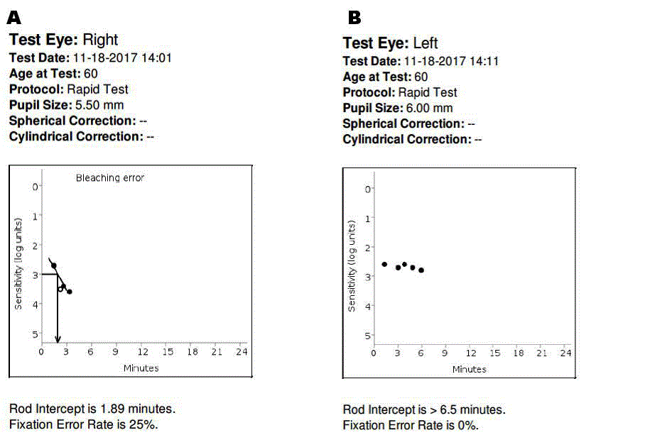
Figure 1
An example of a dark adaptation test performed in the same patient. Figure 1A shows a normal test result performed in the right eye. There is a rodintercept of 1.89 minutes; which is considered normal, a fixation rate of less than 30% is also considered normal. Figure 1B shows the left eye of the same patient, in which the rod-intercept was > than 6 minutes, clinically the left eye showed damage consistent with moderate macular degeneration.
AdaptDx (MacuLogix, Hummelstown, PA)
To perform the test, the patient’s eyes are dilated to 6 mm by using 1% tropicamide and 2.5% phenylephrine hydrochloride. The patient needs to be refracted prior to the test so that corrective lenses can be used as appropriate for the 30 cm viewing distance to correct for blur. The fellow eye is covered with an eye patch. An Infrared camera displays an image of the eye on the operator control system. The patient’s eye is bleached by exposure to a 505 nm photoflash (0.8-ms duration, 1.8 x 10⁴ scotoma cd/m2 s intensity). This is equivalent to 76% bleaching level for rods. The patient has 2 seconds to respond to the stimulus by pushing a response button. For each indication that a stimulus is visible, the intensity is decreased for each successive presentation in steps of 0.3 log units until there are no more responses. At this point the target intensity is increased in 0.1 log units until the patient responds again. This intensity is defined as a threshold. Successive threshold measurements start with the stimulus intensity at 0.2 log units brighter than the previous threshold measurement (Figure 1).
Conclusion
Night vision and low illuminance vision encountered in early AMD is a clinically significant problem similar to visual acuity impairment encountered in late-stage AMD. Dark adaptation is a suitable primary endpoint that aids in the evaluation of treatment efficacy aimed at AMD management.
References
- Christoforidis JB, Tecce N, Dell'Omo R, Mastropasqua R, Verolino M, Costagliola C. Age related macular degeneration and visual disability. Curr Drug Targets. 2011; 12: 221-233.
- Tolentino MJ, Dennrick A, John E, Tolentino MS. Drugs in Phase II clinical trials for the treatment of age-related macular degeneration. Expert Opin Investig Drugs.2015; 24: 183-199.
- Laíns I, Miller JB, Park DH, Tsikata E, Davoudi S, Rahmani S, et al. Structural Changes Associated with Delayed Dark Adaptation in Age-Related Macular Degeneration. Ophthalmology.2017; 124: 1340-1352.
- Argon laser photocoagulation for senile macular degeneration. Results of a randomized clinical trial. Arch Ophthalmol.1982100: 912-918.
- Cervantes-Castaneda RA, Banin E, Hemo I, Shpigel M, Averbukh E, Chowers I. Lack of benefit of early awareness to age-related macular degeneration. Eye (Lond).2008; 22: 777-781.
- Friedburg C, Sharpe LT, Beuel S, Zrenner E. A computer-controlled system for measuring dark adaptation and other psychophysical functions. Graefes Arch Clin Exp Ophthalmol.1998; 236: 31.
- Laíns I, Miller JB, Mukai R, Mach S, Vavvas D, Kim IK, et al. Health Conditions Linked to Age-Related Macular Degeneration Associated with Dark Adaptation. Retina.2017.
- Jackson GR, Clark ME, Scott IU, Walter LE, Quillen DA, Brigell MG. Twelve-month natural history of dark adaptation in patients with AMD. Optom Vis Sci.2014; 91: 925-931.
- Jackson GR, Scott IU, Kim IK, Quillen DA, Iannaccone A, Edwards JG. Diagnostic sensitivity and specificity of dark adaptometry for detection of age-related macular degeneration. Invest Ophthalmol Vis Sci.2014; 55: 1427-1431.
- Jackson GR, Edwards JG. A short-duration dark adaptation protocol for assessment of age-related maculopathy. J Ocul Biol Dis Infor.2008; 1: 7-11.