Case Report
Unilateral Condylar Hyperplasia: A Case Report
Kalyani Gelada*, Rajshekhar Halli, Manjula Hebbale, Husain Mograwala, Sanjana Sethi and Viren S Patil
Department of Oral and Maxillofacial Surgery, Bharati Vidyapeeth Dental College and Hospital, India
*Corresponding author: Kalyani Gelada, B 101, Sujay Garden Housing Society, Mukund Nagar, Pune 411037, India
Published: 20 Jun, 2018
Cite this article as: Gelada K, Halli R, Hebbale M,
Mograwala H, Sethi S, Patil VS.
Unilateral Condylar Hyperplasia: A
Case Report. Ann Clin Case Rep. 2018;
3: 1524.
Abstract
Condylar Hyperplasia (CH) is a bone disease characterized by the increased development of
mandibu¬lar condyle. It regularly presents as an active growth with facial asymmetry generally
without pain. Statistically it af¬fects more women in adolescence, although it does not discriminate
by age or gender. Its best-known consequence is Asymmetric Facial Deformity (AFD), combined
with alteration of the dental occlusion with unilateral crossbite or open bite. It is not known when
condylar hyperplasia begins and how long it lasts. We report a unilateral condylar hyperplasia in 35
year old female managed successfully by surgical intervention.
Keywords: Deviated mouth opening; Hyperplasia; Overgrowth of the mandible; Unilateral
crossbite
Introduction
Overdevelopment of mandible was described by Robert Adams in 1836 who related it to
condylar hyperplasia. Condylar hyperplasia of the mandible is an uncommon idiopathic disorder
of non-neoplastic origin of the jaw characterised by increased volume of the condyle, unilaterally
or bilaterally, leading to facial asymmetry, mandibular deviation, malocclusion and articular
dysfunction generally without pain [1].
Hugo Obwegeser and Makek classified condylar hyperplasia as Type 1, 2, and 3. Wolford et
al developed an updated classification of condylar hyperplasia in 2014 [2] (Table 1). Condylar
hyperplasia has an unknown etiology. Several theories exist in the literature in which one states
that an event of a trauma leading to increase in number of repair mechanism and hormones in
that area may lead to increase in growth of mandible on that side [3]. Another theory states that an
increase in loading of Temporomandibular joint can lead to increase in expression of bone forming
molecules [4]. Condylar hyperplasia predominantly affects women 64% of patients being women
[2]. We report a unilateral condylar hyperplasia in 35 year old female managed successfully by
surgical intervention.
Case Presentation
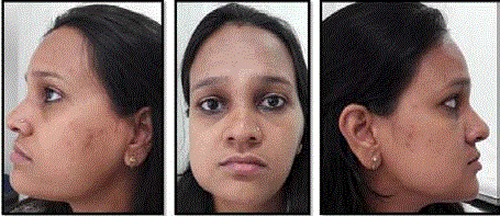
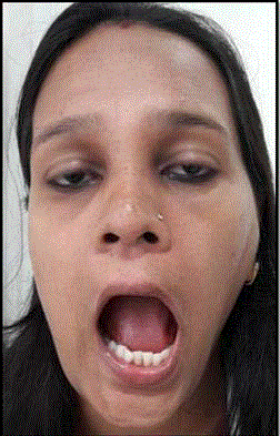
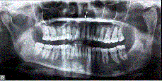
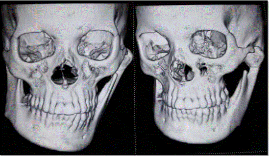
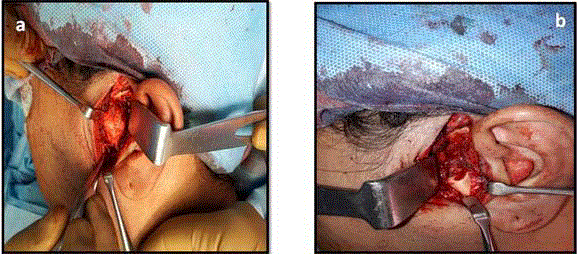
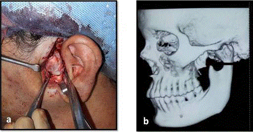
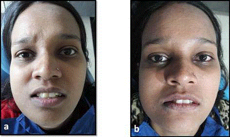
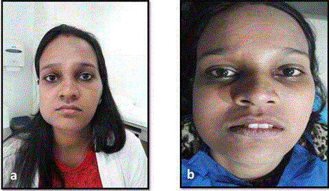
A 35 year old female patient reported to us with a chief complaint of facial asymmetry. Her antenatal and obstetric history was not contributory. She had a history of trauma to the lower jaw during childhood for which she had not undergone any treatment. On extra-oral examination patient had facial asymmetry, flattening seen on the left side of the face with appearance of fullness on right side of the face (Figure 1). A bony swelling in the left pre-auricular region was evident measuring about 1.5 cms x1.5 cms. On opening of the mouth patient had deviation of the lower jaw towards the right side (Figure 2). On radiographic examination, OPG shows gross enlargement of the left condyle and Loss of antegonial notching with downward bowing of the inferior border on the mandible (Figure 3). CT scan revealed a hyperplastic left condyle with elongated neck (Figure 4). An outward and downward growth of the body and ramus of the mandible on the affected side, deviation of mandible and chin to the opposite side, slanted occlusal plane for dental compensation, and deviation of dental midlines were evident. There was asymmetry of the left hemi mandible and outward protrusion of the anterior teeth. As the hyperplastic mass of condyle was evidently extended in the lateral preauricular region, it was easier to approach with preauricular incision (Figure 5a and b). Dissection was carried out till the mass was reached and continued to expose the condylar neck. Bony cut was made just below the hyperplastic mass which led to its separation from the neck of the condyle (Fig 6a and b). Entire mass was carefully removed in Toto (Figure 7a and b). The raw condyle neck portion was rounded off to mimic the condylar head (Figure 5b). Wounds were closed in layers. Post operatively, patient developed open bite on contra lateral side with premature contact on ipsilateral side (Figure 8a and b). Arch bars were placed in both the arches under LA and elastic tractions were initiated till the satisfactory occlusion was obtained for about 3 weeks. The arch bar and elastics were removed once the occlusion was satisfactorily achieved. Patient’s mandibular deviation was appreciably reduced. Patient is on a regular follow-up and there is no occlusal derangement and reduction in the mouth opening noticed (Figure 9a and b).
Figure 1
Figure 1
Pre-operative profile photographs of the patient. On extra-oral
examination patient had facial asymmetry, flattening seen on the left side of
the face and a bony swelling in the left pre-auricular region.
Figure 2
Figure 3
Figure 3
OPG reveals gross enlargement of the left condyle and loss of
antegonial notching with downward bowing of the inferior border on the
mandible.
Figure 4
Figure 5
Figure 5
a) Approach taken with preauricular incision, hyperplastic mass of
condyle was evident. b) Condylectomy followed by roundening of condylar
neck.
Figure 6
Figure 6
a) Bony cut taken on the neck of the condyle, operative site
exposed with preauricular incision. b) Diagrammatic representation of the
surgery performed.
Figure 7
Figure 8
Figure 9
Figure 10
Figure 10
Image obtained by SPECT where a greater capture of the isotope
is observed in the left condyle.
Table 1
Table 1
Hugo Obwegeser and Makek classified condylar hyperplasia as Type 1, 2, and 3. Wolford et al. developed an updated classification of condylar hyperplasia in 2014.
Table 2
Table 2
Various authors suggested different treatment modalities depending on the age and SPECT findings, including condylectomy, compensatory orthodontics and surgical cosmetic camouflage either alone or in combination.
Discussion
The diagnosis of Condylar hyperplasia is essentially clinical; there
are supporting studies that determine the activity and morphology of
the condyle affected [5]. Without a doubt, computerized tomography
(CT) has contributed to establishing the pathology and condylar
morphology but the nuclear medicine studies have helped in
determining the active growth of the condyle. Technetium-99m is
administered with methylene diphosphonate, which is absorbed by
hydroxyapatite crystals and calcium from the bone tissue so that the
fixation intensity is proportional to the degree of osteoblast activity;
the examination that obtains the scanned bone is called “single
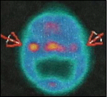
photon emission computed tomography” (SPECT) [6] (Figure 10).
As the degree of asymmetry increases, the subjects determine a
greater need for surgery that repairs esthetics and function, indicating that when the chin asymmetry deviates 10 mm from the midline,
there is a high demand to correct it surgically; this demand decreases
in proportion to increase in the patient’s age and to decrease in the
perception of facial esthetics [7]. Various authors suggested different
treatment modalities depending on the age and SPECT findings,
including condylectomy, compensatory orthodontics and surgical
cosmetic camouflage either alone or in combination [6] (Table 2).
In our case the probable cause was trauma during childhood.
CT and SPECT have contributed largely in determining the time
of surgery. Surgical correction with proper planning and timing of
treatment yields good functional and esthetic outcome.
References
- Raymond Fonseca. Oral and maxillofacial Surgery. 2017; 3: 2696.
- Peter Ward Booth. Oral and Maxillofacial Surgery. 2
- Egyedi P. Aetiology of condylar hyperplasia. Aust Dent J. 1969;14:12-17.
- Fragiskos. Oral and Maxillofacial Surgery.
- Peterson. Principles of Oral and Maxillofacial Surgery.
- Saridin C, Gilijamse M, Kuik D, te Veldhuis E, Tuinzing D, Lobbezoo F et al. Evaluation of temporomandibular function after high partial condilectomy because of unilateral condylar hyperactivity. J Oral Maxillofac Surg. 2010; 68: 1094-1099.
- M Portelli, Giovanni Matarese, Elda Gatto, Alessandra Lucchese. Unilateral condylar hyperplasia: Diagnosis, clinical aspects and operative treatment. A case report. Eur J Paediatr Dent. 2015; 16:
- Wolford LM, Movahed R, Perez DE. A classification sytem conditions causing condylar hyperplasia. J Oral Maxillofac Surg. 2014;72;567-595.

