Case Report
A Rare Cause of Altered Mental Status: The Morel–lavallée Syndrome
Sarıcıcek BM1, Aydın OF1, Bıcakcı S1, Gul E2 and Guneysel O*1
1Lutfi Kirdar Kartal Education and Research Hospital, Emergency Medicine Clinic, Istanbul, Turkey
2Elaziğ Education and Research Hospital, Emergency Medicine Clinic, Elaziğ, Turkey
*Corresponding author: Ozlem Guneysel, Lutfi Kirdar Kartal Education and Research Hospital, Emergency Medicine Clinic, Semsi Denizer Cad. E-5 Karayolu Cevizli Mevkii 34890 Kartal / Istanbul, Turkey
Published: 07 Jun, 2016
Cite this article as: Sarıcıcek BM, Aydın OF, Bıcakcı S,
Gul E, Guneysel O. A Rare Cause of
Altered Mental Status: The Morel–
lavallée Syndrome. Ann Clin Case Rep.
2016; 1: 1013.
Abstract
Altered mental status is a common clinical condition with a wide andvaried diagnosis. Morel– LavalléeSyndrome is rare but may be caused by a serious hemorrhage concluding with hemorrhagic shock. Hemorrhagic shock has occured with altered mental status in a 76 year-old male patient with Morel-Lavallée Syndrome. Altered mental status occured after hemorrhagic shock. The syndrome caused by post-traumatics hearing of the hypodermis from the underlying fascia. Physical examination, MRI and USG is the mainstay of diagnosis and treatment includes both surgical and minimally invasive modalities.
Introduction
Morel-Lavallée Syndrome is defined as the separation of skin and subcutaneous tissues from
underlying fascia and results with a closed space in which haematoma and liquified fat tissues
accumulate [1,2]. Morel-Lavallée Syndrome frequently appears as fluctuating fluid accumulation
[1,2].The lesion most frequently occurs in the greater trochanter, lowerback, knee and scapula [3].
Usually this is a sub cutaneous hematoma, but thehematoma can become so large that it seriously
threatens hemorrhagic shock and the viability of the skin above [2]. Morel–Lavallée Syndrome
is rare but may cause serious hemorrhage which can lend to a rapid reduction in blood volume
[4]. Acute delirium, restlessness, disorientation, confusion and coma are caused by a decrease in
cerebral perfusion pressure [5].
Here were port a case of Morel–Lavallée Syndrome that resulted in hemorrhagic shock with
altered mental status.
Case Report
A 76 year old male presented with the acute on set of altered mental status. Family members
reported that the patient was being less interactive and more anxious than usual. Vital sign sat
the time of admission were as follows; heart rate: 97/min, respiratory rate: 20/min, blood pressure:
140/90 mm- Hg and body temperature: 35.9oC. The blood glucose level was 320 mg/dl.
He was conscious and cooperated but was disoriented. His Glas gow Coma Scale score was
15. He was anxious and fidgety. Extremity examination revealed an ecchymosis along the lateral
right femoral region, a soft, fluctuant mass just inferior to the ecchymosis and a short leg splint. No
laceration or ulceration was present. Bullous lesions were present on the ecchymotic areas (Figure
1). The other system examinations were normal. Normal saline was the fluid of choice for initial
therapy.
The patient was hospitalized for a talus fracture caused by the traffic accident 2 days before.
Talus fracture was treated with a short leg splint. After two days, he was discharged and prescribed
enoksaparin with an additional follow-up observation. Past medical history revealed that type-2
diabetes mellitus, chronic obstructive pulmonary disease and hypertension were present. His
medication was β blocker.
Laboratory results were as follows; hemoglobin: 7.7g/dl, hematocrit: 22.5 (2 days before
his hemoglobin level was 13.4 g/dl, hematocrit was 39.3), glucose: 336 mg/dl. Two units RBC
preparations were immediately transfused. After transfusion, laboratory results were as follows:
hemoglobin: 9 g/dl, hematocrit: 26.5. As soon as hemodynamic status had stabilized, thorax and
abdominal computer tomography scans were performed. There were no pathologic findings.
The diagnosis was confirmed with magnetic resonance (MRI)
which revealed that a well- marginated, T2 hyperintense, lentiform
collection, between the sub cutaneous fat and fascia (Figure 2). The
lesion measured 45 mm in dimension. The marrow signal of adjacent
femur was normal. Surrounding muscular edema, sinus tracts or
cutaneous defects were not present.
The patient was admitted to orthopedic surgery ward. The fracture
was stabilized with a short leg splint. The hematoma was drained with
a small incision and a compression bandage was applied. Damaged
tissue debridement was performed after the patient was stabilized. A
partial-thickness graft was applied. A drain was placed and delayed of
a day. On the fifth day he was discharged with an additional follow-up
observation.
Figure 1
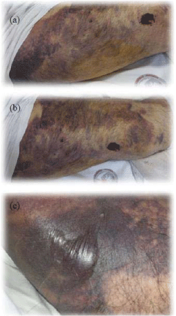
Figure 1
a) An ecchymosis along the lateral right femoral region. b) A soft, fluctuant mass just inferior to the ecchymosis. c) Bullous lesions were present on the ecchymotic areas.
Figure 2
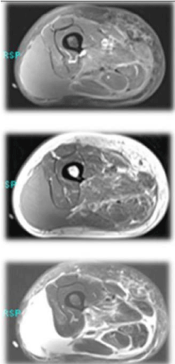
Figure 2
Well-marginated, T2 hyperintense, lenti form collection was
present between the sub cutaneous fat and fascia.
Discussion
Morel- Lavallee Syndrome frequently appears as a fluctuating fluid
accumulations [1]. In a multitrauma patient whose hemodynamic
status was stabilised, a physical examination should be performed
on an undressed patient and should begin with a quick head-to-toe
inspection and all skin lesions on the whole body should be evaluated
carefully for the diagnosis [1]. A fluctuating lesion and excessive
skin mobility with manual examination was accepted as the finding.
MRI and ultrasonography have been recommended to confirm the
diagnosis [2].
Variable treatment options have been suggested for MorelLavallée
syndrome. Wound drainage and a compression bandage was applied. Non-invasive treatment was performed with elastic bandages
in closed degloving skin lesions [1]. The primary modality is to treat
the soft-tissue problem at the same time the fracture is stabilized
[2]. All of the treatment options have many complications, such as
recurrence of the swelling, increased incidence of infection, skin
necrosis etc.
Conclusion
Morel–Lavallée Syndrome is a rare cause of sub cutaneous
swelling. Some times the disease can not be identified and may be
fatal. Clinicians should be careful of the probable diagnosis.
Acknowledgement
I would like to thank Stephen Mason who were generous with his time and aid.
References
- Harma A, Inan M, Ertem K. The Morel-Lavalleelesion: a conservative approach to closed degloving injuries. Acta Orthop Traumatol Turc. 2004; 38: 270-273.
- Wood GW. General Principle of Fracture Treatment. Canale ST, Beaty JH.Campbell's Operative Orthopaedics. Philadelphia: Mosby Elsevier. 2008; 3018-3080.
- Moriarty JM, Borrero CG, Kavanagh EC. A rare cause of calf swelling: the Morel-Lavallee syndrome. Ir J MedSci. 2011; 180: 265-268.
- Jones AE, Kline JA. Shock. In: Marx JA (ed) Rosen's Emergency Medicine. MosbyElsevier, Philadelphia. 2010; pp 34-41.
- Otero RM. Approach to the Patient in Shock. Tintinalli EJ.Tintinalli's Emergency Medicine. New York: McGrawHill. 2011; 165-172.