Case Report
Intrapleural Fat-Fluid Level: A Unique Sign in Chest Imaging
Barry Hutchinson*
Department of Radiology, NYU School of Medicine, USA
*Corresponding author: Barry Hutchinson, NYU School of Medicine, Bellevue Medical Center, Department of Radiology, 462 First Avenue, New York, NY 10016, USA
Published: 01 Feb, 2018
Cite this article as: Hutchinson B. Intrapleural Fat-Fluid
Level: A Unique Sign in Chest Imaging.
Ann Clin Case Rep. 2018; 3: 1500.
Abstract
Pseudochylothorax (PCT) describes the accumulation of a lipid-rich pleural effusion that resembles a chylothorax at thoracocentesis but that does not result from obstruction of the thoracic duct. They most commonly occur in the setting of a chronic tuberculous empyema. On Computed Tomography (CT) PCT may be indistinguishable from simple pleural effusions but in rare cases they can contain macroscopic fat that forms either a fat-fluid of fat-calcium level. This unusual appearance can pose a diagnostic dilemma to the radiologist unfamiliar with this entity. We present two such cases with CT imaging.
Case Presentation
Case 1
A 28 year-old man with on an anti-tuberculosis regimen presented with a 4 month history of
increasing fever and breathlessness. His history was significant for a spontaneous pneumothorax
8 months prior for which he had undergone a thoracoscopic bleb resection. One month later he
developed unexplained severe thrombocytopenia for which a splenectomy was performed. At
histology the spleen was virtually replaced by caseating granulomas and an Acid-Fat Bacillus (AFB)
smear was positive. The patient was initiated on tuberculosis treatment. A chest radiograph on
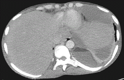
admission (not shown) revealed a loculated left pleural effusion. A CT scan performed showed a
posteriorly loculated pleural fluid collection (Figure 1) containing fat with a density of -45 Hounsfield
Units (HU). Fat layered posteriorly forming a fat-fluid level with an anterior fluid collection with a
mildly increased density of 25 HU suggestive for complex fluid. A complex fluid collection without
macroscopic fat was seen loculated anteriorly. Mild pleural thickening was present but there was
no visible pleural calcification present. Percutaneous drainage of the more posterior fluid revealed
non-purulent fluid that resembled milk. Fat analysis of this fluid revealed a triglyceride level of
134 mg/dL. White blood cells were increased with 89% lymphocytes. The pleural collections were
negative at AFB smear and at Gram stain and cultures for both mycobacterial and other microbes
were negative. The effusion was subsequently drained using Video-Assisted Thoracoscopic Surgery
(VATS). The patient had a prolonged inpatient post-operative course complicated by diffuse
endobronchial spread of tuberculosis but was eventually discharged well with a diagnosis of infected
PCT secondary to tuberculous empyema.
Case 2
A 57 year-old malewith an unknown history underwent a routine chest radiograph that revealed
a rounded pleural-based opacity arising from the right lateral chest wall (not shown). A CT showed
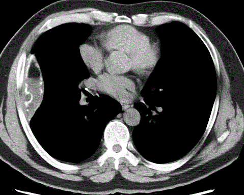
a loculated fluid collection within the right lateral pleural space with calcifications and a fat-fluid
level with fat (-115 HU) occupying the non-dependent portion of the collection (Figure 2). The lungs were clear with no imaging evidence for active tuberculosis. As the patient was asymptomatic,
she was managed conservatively. Limited clinical details were available in the follow-up in this case
but imaging appearances are typical for a PCT.
Figure 1
Figure 2
Figure 2
A CT revealed a locualted right pleural collection with calcifications
and a fat-fluid level with fat (-115 HU) occupying the non-dependent portion
of the loculated collection.
Discussion
A pleural effusion with a milky or turbid appearance on thoracocentesis has been associated
with a limited number of diagnoses including chylothorax and pseudochylothorax (PCT) [1]. A
chylothorax describes leakage of chyle into the pleural space usually secondary to obstruction of
the thoracic duct [1,2]. The terms pseudochylous pleural effusion, chyliform pleural effusion and cholesterol effusion have all been used to describe a pleural effusion that is not truly chylous in
nature but with very high lipid content and can be applied to a variety of etiologies [3]. They are usually associated with a chronic pleural effusion and chronic pleural inflammation [2]. Distinguishing between these two entities
is important for determination of etiology and for management
planning [1].
A PCTis formed within a chronic pleural effusion usually
surrounded by thickened and fibrotic pleurae [3]. The typical patient
is male, aged between 40 to 75 [4]. Approximately one third of
patients with pseudochylothorax will be asymptomatic. Symptoms
when present are usually related to the restrictive respiratory
defect [1]. These pleural effusions are usually long-standing with a
mean duration of 5 years prior to development of visible lipid [2,5].
Development of fat within the effusion is thought to represent a step
in the maturation of a chronic effusion rather than an acute process
[3] and the identification of PCT can be considered a form of lung
entrapment in the setting of chronic inflammation [1]. The exact
pathogenesis of the PCT remains uncertain. Any long-standing
pleural effusion tends to become populated by lymphocytes [2] and a
common theory is that fat is generated from the degeneration of white
and red blood cells in the pleural space. Reduced transfer of lipids out
of the pleural space due to pleural fibrosis has been suggested as the
reason the large quantity of lipid within the pleural collection [3,4].
Synthesis of fat by the pleura has also been postulated [1]. The large
majority of reported cases of PCT have mainly described either as a
sequela of tuberculous pleural effusion or in the setting of rheumatoid
arthritis. In one review, 88.5% of cases were secondary to one of
these two etiologies [4]. However PCT’s have also been identified in
the setting of trauma includingpost-operative patients, pulmonary
embolism, pulmonary Echinococcal and Paragonimiasis infection
among others [3,4,6,7].
Despite their high lipid content, both chylothoraces and PCT’s
are highly variable in their attenuation CT [8] due to their high
protein content and sometimes hemorrhagic components [4,9].
Therefore, the diagnosis of pseudochylothorax is usually made at
thoracocentesis. Chylothoraces and PCT’s can be discriminated
using lipid analysis of the aspirated pleural fluid [2]. PCT’s are usually
associated with significantly elevated cholesterol concentrations [1]
and the presence of cholesterol crystals is diagnostic. Triglyceride
concentrations, while increased, are usually considerably lower
in PCT’s when compared with chylothoraces and a cholesterol to
triglyceride ratio of >1 is virtually always present [4]. The presence
of chylomicrons is not seen in PCT’s and confirms the diagnosis of
chylothorax [1,2].
While PCT’s are common in the setting of a pleural peel, [10] visualization of macroscopic fat (-90 to -115 HU) layering inthe nondependent aspect of the fluid collection or adjacent to calcification
(fat-calcium level) on Computed Tomography (CT) is extremely
rare having only been described in a single case series of 6 patients
[3]. The presence of a fat-fluid level in a pleural effusion is almost
exclusive to PCT, only having beendescribed in a single case report
of a mediastinal teratoma rupturing into the pleural space [11]. It
has not been reported in the setting of a true chylothorax. Given that
the effusions are most commonly tuberculous, loculation is usually
present [9]. Chyle is non-inflammatory and does not injure the
pleural membranes; this may help to distinguish the 2 entities in the
absence of macroscopic fat [1].
They are often large with 96.3% occupying > 1/3 of the
hemithorax [4]. Pleural thickening is not necessary for diagnosis
but is usually seen, observed in up to 80% of such case, in a recent
review [4]. Most patients have a remote history of pleurisy. In the
cases series by Song et al. 5 of 6 patients had a documented history
of tuberculous pleural effusion. All of these patients demonstrated
pleural thickening (4-10 mm) and all had calcification. Bacteriological
confirmation of tuberculous etiology is often limited in aspirates
of conventional tuberculous pleural effusions and appears to be
even more so in pseudochylothoraces [9]. Decortication and/or
pleuropneumonectomy was performed in 4 of the 5 patients with
a history of tuberculous effusion from the above series. Histology
revealed chronic caseating granulomatous inflammation with fibrosis
at histological but Mycobacteria species could not be confirmed at
staining or culture.
Management of pseudochylothoraces is not well-established [4].
While successful decortication with or without pleurectomy was
performed in 4 of the 5 patients with tuberculous pleural effusions
in Song’s cohort; these patients all had symptoms related to their
pleural effusions [3]. Both favorable and unfavorable outcomes have
been seen with antimycobacterial chemotherapy, decortication,
pleurectomy and pleurodesis in patients with chronic tuberculous
pseudochylothoraces [4]. Many authors advise intervention only in
the setting of symptoms or an increasing effusion [10].
Conclusion
The presence of a milky-appearing effusion in the presence of visualized pleural thickening or calcification should alert a clinician to the diagnosis [1]. While uncommon, the presence of a fat/fluid level within a pleural effusion with pleural thickening on CT has only been described in pseudochylothoraces and given its rarity could be misdiagnosed as a fat-containing pleural mass by the radiologist unfamiliar with this diagnosis.
References
- Agrawal V, S Sahn. Lipid pleural effusions. Am J Med Sci. 2008; 335: 16-20.
- Hooper C, YG Lee, N Maskell. Investigation of a unilateral pleural effusion in adults: British Thoracic Society pleural disease guideline 2010. Thorax. 2010; 65: ii4-ii17.
- Song JW, Im JG, Goo JM, Kim HY, Song CS, Lee JS, et al. Pseudochylous Pleural Effusion with Fat-Fluid Levels: Report of Six Cases 1. Radiology. 2000; 216: 478-480.
- Adriana Lama, Lucía Ferreiro, María E Toubes, Antonio Golpe, Francisco Gude, José M Álvarez-Dobaño, et al. Characteristics of patients with pseudochylothorax—a systematic review. J Thorac Dis. 2016; 8: 2093-2101.
- Choi WS, Park CM, Song YS, Lee SM, Wi JY, Goo JM. Transient subsolid nodules in patients with extrapulmonary malignancies: their frequency and differential features. Acta Radiol. 2015; 56: 428-37.
- Crina Muresan, Lucian Muresan, Ioana Grigorescu, Dan L Dumitrascu. Chyliform effusion without pleural thickening in a patient with rheumatoid arthritis: A case report. Lung India. 2015; 32: 616-619.
- Carel RS, G Schey, I. Bruderman. Chyliform Pleural Effusion: An Unusual Manifestation of Hepatothoracic Echinococcus Cysts. Chest. 1975; 68: 598-599.
- Kim EA, Lee KS, Shim YM, Kim J, Kim K, Kim TS, et al., Radiographic and CT Findings in Complications Following Pulmonary Resection 1. Radiographics. 2002; 22: 67-86.
- Jolobe OM. Atypical tuberculous pleural effusions. Eur J Intern Med. 2011. 22: 456-459.
- Hillerdal G. Chylothorax and pseudochylothorax. Eur Respir J. 1997; 10: 1157-1162.
- Yeoman LJ, HR Dalton, EJ Adam. Fat-fluid level in pleural effusion as a complication of a mediastinal dermoid: CT characteristics. J Comput Assist Tomogr. 1990; 14: 307-309.


