Case Report
Propionibacterium acnes Infection of a Mitral Annuloplasty Ring
Valentino MA1*, Saxena S2, Vira A2 and Tecce M1
1Division of Cardiology, Department of Internal Medicine, Thomas Jefferson University Hospital, Philadelphia, USA
2Department of Internal Medicine, Thomas Jefferson University Hospital, Philadelphia, USA
*Corresponding author: Valentino MA, Division of Cardiology, Department of Internal Medicine, Thomas Jefferson University Hospital, Philadelphia, PA, USA
Published: 24 Apr, 2017
Cite this article as: Valentino MA, Saxena S, Vira A, Tecce
M. Propionibacterium acnes Infection
of a Mitral Annuloplasty Ring. Ann Clin
Case Rep. 2017; 2: 1343.
Abstract
Eosinophilic pustular folliculitis (EPF) is a rare, idiopathic, recurrent, self-limiting disease. It presents as sterile papulopustules located on the scalp and sometimes also on other body parts, without signs of systemic illness. Ten percent of cases occur in infants, mainly before the age of 6 months. The etiology is unknown. Eosinophilic pustular folliculitis of infancy (EPFI) is often underdiagnosed and overtreated, especially in a vulnerable population like preterm infants. We present a female neonate, born prematurely at a gestational age of 25 weeks. She developed recurrent papulopustules on an erythematous base with crustae. After initial treatment with antibiotics and antifungal agents, she was diagnosed with EPFI. We discuss the differential diagnosis and management of EPFI and its possible relationship with the immunological immaturity associated with preterm birth.
Keywords: Eosinophilic pustular folliculitis; Newborn; Infant; Premature; Hyper-IgE syndrome
Introduction
Eosinophilic pustular folliculitis (EPF), also known as Ofuji disease, is a rare, idiopathic,
inflammatory skin disease. Eosinophilic pustular folliculitis of infancy (EPFI) accounts for 10% of
all EPF cases. It occurs four times more often in (Caucasian) boys than girls [1,2]. The timing of
first presentation ranges from birth to 1.5 years; 70% presents before the age of 6 months [2]. It is
a recurrent disease in all children with outbreaks every 1-12 weeks, each lasting 1-4 weeks [1,2]. In
80%, the disease spontaneously resolves around the age of 3 years [2].
At presentation, infants have sterile papulopustules on an erythematous base. The lesions are
always located on the scalp and in 65% also on other body parts, without signs of systemic illness.
Lesions form groups, sometimes with secondary crusting. Unlike adult eosinophilic folliculitis,
lesions lack the typical halo [1,3]. After remission, local hyperpigmentation can be seen. Recurrences are often located at hyperpigmented old lesion sites.
Laboratory investigations reveal blood eosinophilia in up to 83% of cases. This might be an
underestimation, since blood eosinophilia is only present in the acute phase of the outbreak [2,4]. Microbiology shows negative cultures for bacteria, fungi, and viruses although secondary infection
with Staphylococcus aureus might occur [1]. At a young age, it may be difficult to differentiate
between EPFI and hyper-IgE syndrome (HIES, Job syndrome), since both are accompanied by
papulopustular dermatitis, often located on the scalp and face, with crustae associated with S.
aureus and folliculitis with eosinophilic infiltrates. Both disease entities feature blood eosinophilia.
Moreover, elevated IgE levels may be absent in infants with HIES [5].
Histology in EPFI reveals folliculitis in only 62% of cases whereas folliculitis is obligatory in
the adult counterpart [1-3]. Punch biopsy of the skin shows dense eosinophilic infiltrates in most patients. These are located in the dermis and can be arranged in a peri- or interfollicular fashion.
Lymphocytes and histiocytes can also be present [1]. Further histological findings are subcorneal pustules and spongiosis in the epidermis [6,7].
The cause of EPFI is unknown. Hypersensitivity reactions, immunologic, hormonal, and genetic
etiologies have been suggested [3,8,9].
Case Presentation
A female neonate was born prematurely at 25 weeks and 6 days of gestation due to cervical
insufficiency. Shortly after birth, intravenous treatment with fluconazole was initiated and
continued for 14 days because of a generalized erythematous skin rash with a positive skin swab for
Candida albicans. Considering the unexplained prematurity, the skin rash, and a C-reactive protein (CRP) level of 28 mg/l, we also suspected a perinatal infection, for
which a 7-day course of antibiotics (amoxicillin and gentamicin)
was prescribed. During the first 2 days of her life, she was ventilated
and treated with surfactant because of infant respiratory distress
syndrome (IRDS). No corticosteroids were administered. Her father
was allergic to cats, dogs, dust mite and several fruits and vegetables
in his childhood. These allergies resolved within 2 years. There was no
family history of similar skin lesions or recurrent infections.
At the age of 5 weeks, she developed progressive erythematous
papulopustules with crustae on the scalp, cheeks, and in the supraauricular
region (Figure 1 and 2), without any signs of systemic
inflammation. Routine laboratory investigations, including
leukocytes and CRP, were normal. She was initially treated with
topical fusidic acid and intravenous Flucloxacillin and Fluconazole.
These medications were discontinued after a week when blood
cultures and regular skin swabs turned out negative for bacteria and
fungi.
Two weeks later, new lesions appeared on the same sites as the old,
healing lesions. Additional lesions emerged on the abdomen. Initially,
no treatment was started. However, because of the progressive nature
of the lesions and the vulnerability of this preterm infant, treatment
with intravenous Flucloxacillin and oral Fluconazole was started
again for a week. This time, a lesion swab showed secondary infection
with S. aureus.
Specialists in paediatric dermatology and paediatric infectious
diseases and immunology were consulted. A differential diagnosis
was made, mainly comprising EPFI and HIES. Additional
laboratory investigations were carried out. Leukocyte differentiation
demonstrated 17% eosinophils (normal <6%), 23% neutrophils
(normal 15%-60%), and 46% lymphocytes (normal 12%-68%).
The absolute blood eosinophil count was 2.78x10^9/l (normal
<0.6x10^9/l). The serum IgE level was <2 U/ml (normal <2 U/l) and
a dihydrorhodamine (DHR) flow cytometric assay (i.e. granulocyte
function test) to assess oxidative burst in our patient’s neutrophils
was normal.
To confirm our clinical suspicion of EPFI, a punch biopsy from a
scalp lesion was performed. This revealed a superficial dermal infiltrate
with a preponderance of eosinophils located in and around a follicle
with follicular destruction (Figure 3). The epidermal epithelium
showed spongiosis with eosinophilic and neutrophilic infiltration.
Periodic-Acid Schiff (PAS) staining was negative for fungal infection.
We diagnosed our patient with EPFI. Her further clinical course
was uneventful. At this moment, she is 4 months old and thriving at
home. Recurrences are becoming less elaborate and less frequent. The
absolute blood eosinophil count decreased to 1.04x10^9/l. Outpatient
follow-up with determination of the serum IgE level at the age of 1
year is planned to rule out HIES.
Figure 1
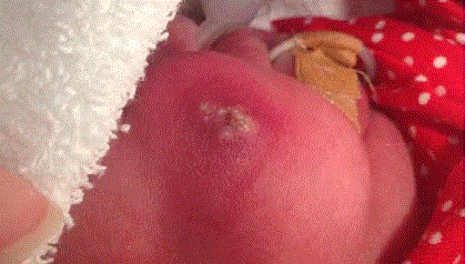
Figure 1
Patient’s right cheek, showing confluent papulopustules on an
erythematous base.
Figure 2
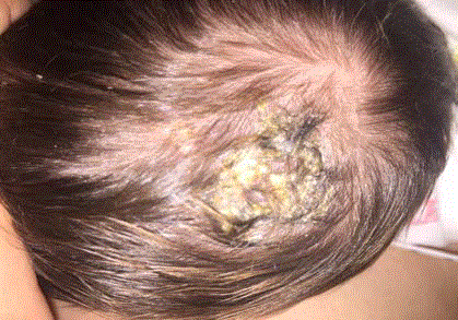
Figure 2
Patient’s scalp with crops of papulopustules and crustae.
Figure 3
Figure 3
Haematoxylin-eosin stain, close up. Dermis and hair follicle
infiltrated by many eosinophilic granulocytes.
Discussion
We describe an extremely preterm female infant having
recurrent eruptions of erythematous papulopustules with crustae,
predominantly located on the face and scalp, starting at the age
of 5 weeks. Although clinical and biochemical signs of systemic
inflammation were absent, she was treated twice with antibiotic
and antifungal medication, without a clear response. Eventually,
the age of onset, appearance, typical localization, and the recurrent
and self-limiting nature of the eruption, in combination with blood
eosinophilia and lack of systemic inflammation, led to a diagnosis of
EPFI, which was confirmed with a skin biopsy.
Primary infectious causes of papulopustular lesions in newborns,
such as bacterial (impetigo, listeriosis, pustular folliculitis), viral
(herpes simplex, varicella), fungal (Candida albicans, malessezia),
and parasitic (in older patients scabies) infections should always be
considered and, if likely, ruled out by appropriate microbiological
investigations [1-3,6,8-10].
The differential diagnosis of noninfectious papulopustular lesions
in newborns consists of EPFI, HIES, erythema toxicum neonatorum,
transient neonatal pustular melanosis, infantile acropustulosis,
Langerhans cell histiocytosis, milliaria pustulosa, acne neonatorum,
incontinentia pigmenti, pustular psoriasis, and transient
myeloproliferative disorder [1-3,6-10]. It is especially challenging
to differentiate between EPFI, erythema toxicum neonatorum, infantile acropustulosis, and HIES [1,3,6,10]. These eruptions can
have a rather similar appearance, with lesions situated on the scalp,
and eosinophilic infiltrates seen in histological specimens. Both EPFI
and infantile acropustulosis (IA) are recurrent. However, lesions are
mainly located on the hands and feet in IA. Also, neutrophils, not
eosinophils, are the prevailing leukocytes found in the pustules of
IA. Erythema toxicum neonatorum has an onset before the age of 2
weeks. HIES can hardly be differentiated form EPFI at a young age,
neither clinically nor histologically. Many patients with HIES were
initially diagnosed with EPFI in their early years. Inasmuch as HIES
entails a risk of skeletal abnormalities and severe infections, among
others, children diagnosed with EPFI should be invited for follow-up
[2]. Also because of the positive cultures for Candida albicans and
Staphylococcus aureus, we still acknowledge the possibility of HIES
in our patient.
Despite the unclear etiology of EPFI, there are several speculative
causes, as mentioned above. We believe that an immunological cause
is plausible, since EPFI is frequently found in (extremely) preterm
infants, who lack a properly developed immune system. Often, these
preterm infants have a history of systemic candida infection, [11] as
was the case in our patient. This also implies an increased vulnerability
of immunocompromised patients for EPFI. Furthermore, EPF is seen
in immunocompromised adults with human immunodeficiency
virus (HIV) and malignancies, such as T-cell lymphoma [12].
Treatment of EPFI with topical corticosteroids should only be
given to patients with complaints of pruritus. Antibiotics should be
reserved for patients with secondary infection of the lesions. Other
treatment modalities have not been proven effective and since the
disease is self-limiting, they carry a large risk of side effects and
overtreatment.
In conclusion, EPFI should be considered when newborns or
infants have a papulopustular eruption, especially when it is recurrent,
located on the scalp, and accompanied by blood eosinophilia. Early
recognition may preclude unnecessary treatment, although this
remains difficult considering the vulnerable nature of this population
and the serious differential diagnoses.
References
- Porriño-Bustamante ML, Sánchez-López J, Aneiros-Fernández J, Burkhardt P, Naranjo-Sintes R. Recurrent pustules on an infant’s scalp with neonatal onset. Int J Dermatol. 2016;55:505-508.
- Hernández-Martín A, Nuño-González A, Colmenero I, Torrelo A. Eosinophilic pustular folliculitis of infancy: a series of 15 cases and review of the literature. J Am Acad Dermatol 2012;68:1:150-155
- Darmstadt GL, Tunnessen WW, Sweren JR, Sweren RJ. Eosinophilic Pustular Folliculitis. Pediatrics 1992;89:1095.
- Taïeb A. Infantile eosinophilic pustular folliculitis in infancy: a nonfollicular disease. Pediatr Dermatol 1994;11:186.
- Van Dijk EM, de Vries E. Immunodeficiency: children with elevated IgE. Ned Tijdschr Allergie & Astma 2013;13:108-113.
- Buckley DA, Munn SE, Higgins EM. Neonatal eosinophilic pustular folliculitis. Clinical dermatology 2001;26:251-255.
- Tucker M, Ramolia P, Wells PJ. JAAD Grand Rounds. Neonate with extensive papulovesicles. J Am Acad Dermatol. 2013;68:877-879.
- Boone M, Dangoisse C, André J, Sass U, Song M, Ledoux M. Eosinophilic pustular folliculitis in three atopic children with hypersensitivity to Dermatophagoides pteronyssinus. Dermatology. 1995;190:164-168.
- Asgari M1, Leiferman KM, Piepkorn M, Kuechle MK. Neonatal eosinophilic pustulosis. Int J Dermatol 2006;45:131-134.
- Ghosh S. Neonatal pustular dermatosis: an overview. Indian J Dermatol 2015;60(2):211.
- Finelt N, Kristal L. Patient characteristics of neonatal eosinophilic pustulosis. Pediatr Dermatol. 2013;30: e204-e207.
- Fuhiyama T, Tokura Y. Clinical and histopathological differential diagnosis of eosinophilic pustular folliculitis. J Dermatol. 2013;40:419-423.