Case Report
Copper Deposition in Cornea and the Lens Capsule as an Ocular Manifestation of Multiple Myeloma
Mina Naderi* and Mohsen Sfandbod
Department of Ophthalmology, Tehran University of Medical Sciences, Iran
*Corresponding author: Mina Naderi, Department of Ophthalmology, Tehran University of Medical Sciences, Tehran Province, Tehran, District 6, Poursina Street, Iran
Published: 25 Nov, 2017
Cite this article as: Naderi M, Sfandbod M. Copper
Deposition in Cornea and the Lens
Capsule as an Ocular Manifestation of
Multiple Myeloma. Ann Clin Case Rep.
2017; 2: 1476.
Abstract
A 50 year-old woman presented to the hospital with a complaint of bilateral gradual progressive visual loss since three years ago, associated with cloudy discoloration of cornea in both eyes with copper deposition in the Descemet’s membrane and deep layers of central cornea along with anterior lens capsule showed on Slit-lamp examination with other normal physical examinations. Through the pattern of copper deposition as previous reported other evaluations were around plasma cell disorders such as multiple myeloma. Laboratory evaluations demonstrated hypercupremia and increased ESR and mild anemia and increased monoclonal IgG and free LAMBDA light chain spike which is compatible with multiple myeloma. Ocular manifestations of multiple myeloma are rare and adverse, hence there is no single comprehensive protocol that can be extrapolate to these various dilemmas.
Case Presentation
A 50 year-old woman presented to the hospital with a complaint of bilateral gradual progressive
visual loss for the preceding three years, associated with cloudy discoloration of cornea in both eyes
(Figure 1). Best-corrected visual acuity was 15/20 in both eyes. Pupillary examination and ocular movement were normal.
Slit-lamp examination showed copper deposition (brown/green), diffusely scattered in the
Descemet’s membrane and deep layers of central cornea along with anterior lens capsule. Apart
from this, she had no remarkable past medical history, familial history or exposure to copper.
Laboratory evaluations demonstrated hypercupremia, normal ceruloplasmin levels and rose. ESR:
50 (M: up to 12 mm/hr and F: up to 22 mm/hr)
Upon further assessment, there were not any evidence of distress and she appeared to rest
in a comfortable state. Moreover, her vital signs were well within normal limits. Except for the
ocular changes, physical examinations were unremarkable with no hepatosplenomegaly or palpable
lymphadenopathy.
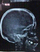
Skull X ray of AP and lateral view showed multiple punched out lesions over skull bone (Figure
2).
In the light of findings through the pattern of copper deposition as previous reported, we
decided to evaluate the patient for suspected plasma cell disorders. The patient underwent a serum
protein electrophoresis, which showed a monoclonal IgG (25.020. nl range: 6.57-18.37) and free
LAMBDA light chain spike (42.1nl range: 5.7-26).
Bone marrow aspiration and biopsy revealed an increased population of abnormal and
dysplastic plasma cell (35% of the cellular elements) which was compatible with the diagnosis of
multiple myeloma. The patient was diagnosed with multiple myeloma based on the laboratory,
histology and imaging findings. The clinical finding of copper deposition in Descemet's membrane
and in the anterior lens capsule associated with in the Descemet’s membrane and deep layers of
central cornea along with anterior lens capsule are consistent with the previous reported patients
with multiple myeloma [1-5] and monoclonal gammopathy of undetermined significance (MGUS)
[6-10]. Although in this case the posterior lens capsule remains intact. The reason behind ocular deposition of copper in multiple myeloma and its unique pattern which differs from Wilson disease
still eludes us. One suggested mechanism for this phenomenon is that IgG λ has an affinity of copper
in multiple myeloma and consequently contribute to the copper transfer into the anterior chamber
and then diffuse and bind to Descemet's membrane [4]. Copper has a special affinity for the
basement membrane and accumulations in Descemet's membrane as we can see the characteristic kayser-fleischer ring in Wilson disease [11].
The patient was treated with Velzomib, dexamethasone and
thalidomide (so called VTD regimen). Moreover D-penicillamin was
added to decrease serum copper level. After completed remission she
was referred to a center for autologous Bone Marrow Transplantation.
Figure 1
Figure 1
Bilateral cloudy cornea due to copper deposition in Descmet's
membrane and anterior capsules.
Figure 2
References
- Goodman SI, Rodgerson DO, Kauffman J. Hypercupremia in a patient with multiple myeloma. J Lab Clin Med. 1967; 70:57-62.
- Lewis RA, Falls HF, Troyer DO. Ocular manifestations of hypercupremia associated with multiple myeloma. Arch Ophthalmol. 1975; 93:1050-1053.
- Hawkins AS, Stein RM, Gaines BI, Deutsch TA. Ocular deposition of cupper associated with multiple myeloma. Am J Ophthalmol. 2001; 131: 257-259.
- Edward DP, Patil AJ, Sugar J, Parikh M. Copper deposition in a variant of multiple myeloma: pathologic changes in the corna and the lens capsule. Cornea. 2011; 30: 360-363.
- Haung YH, Tseng SH. Corneal snowflakes. Lancet. 2012; 380: 506.
- Adave AJ, King JA, kim BT, Hopp L. Corneal copper deposition associated with chronic lymphocytic leukemia. American journal of ophthalmology. 2006; 142:174-176.
- Martin NF, Kincaid MC, Stark WJ, Petty BG, Surer JL, Hirst LW, et al. Ocular cupper deposition associated with pulmonary carcinoma, IgG monoclonal gammopathy and Hypercupremia. A clinicopathologic correlation. Ophthalmology. 1983; 90:110-116.
- Probst LE1, Hoffman E, Cherian MG, Yang J, Feagan B, Adams P, et al. Occular cupper deposition associated with benign monoclonal gammopathy and hypercupremia. Cornea. 1996; 15:94-98.
- Tzelikis PF, Laibson PR, Ribeiro MP, Rapauno CJ, Hammersmith KM, Cohen EJ. Ocular copper deposition associated with monoclonal gammopathy of undetermined significance: case report. Arq Bras Oftalmol. 2005; 68:539-541.
- Shah S, Espana EM, Margo CE. Ocular manifestations of monoclonal copper -binding immunoglobulin. Surv Ophthalmol. 2014; 59:115-123.
- Brewer GJ, Yasbasiyan-Gurkan V. Wilson disease. Medicine. 1992; 71: 139-164.


