Case Report
The Importance of Osteitis as the Originating Site of Inverted Papilloma
Alejandro Martín Vargas-Aguayo*
Department of Otorhinolaryngology, Centro Médico ABC, Mexico
*Corresponding author: Alejandro Martín Vargas Aguayo, Department of Otorhinolaryngology, Centro Médico ABC, Av. Carlos Graef Fernández 154-304, Colonia Tlaxala Santa Fe, Delegación Cuajimalpa, C.P. 05300, Ciudad de México, México, USA
Published: 22 Nov, 2017
Cite this article as: Vargas-Aguayo AM. The Importance
of Osteitis as the Originating Site of
Inverted Papilloma. Ann Clin Case Rep.
2017; 2: 1473.
Abstract
While inverted papilloma is a rare tumor occurring in only 0.5-4 % of all primary nasosinal tumors, its importance lies in the fact that retrospective studies have proven that 2% to 3% of nasal polyps end up being inverted papilloma. Since about 2006, it has been found that the originating site of inverted papilloma in most cases is a zone of osteitis (or hyperostosis) that needs to be detected by means of a computerized tomography. This has resulted in a lower recurrence rate when combined with endoscopic surgery. Below, a case of inverted papilloma with osteitis in the anterior ethmoidal air cells will be presented (Stage Krouse 2).
Keywords: Inverted papilloma; Osteitis; Hyperostosis
Introduction
Inverted papilloma (IP) is a rare, benign tumor, aggressive by nature, that causes local damage, erodes bone [1-3], tends to recur, is multicentric at a rate of 4% and is related to malignancy at a rate of 5% -15% [4]. In the year 2000, Doctor Krouse published his staging report founded on 30 years of experience with IP; this classification was based on the tendency of IP to recur. His main objective is to offer prognostic information [5]. Even before the year 2000, it was shown that radical surgery (medial maxillectomy) of IP lowered its recurrence; the key anatomic area in Krouse’s (and in others’) staging, is the medial wall of the maxillary sinus. If the tumor originates on another maxillary sinus wall that is not the medial wall, or on the frontal or sphenoid sinuses, the possibility of recurrence is greater. This also applies to the resection of IP in the endoscopic age [5,6]. Around 2006, however, the first studies on osteitis (or hyperostosis) as the originating site of IP, which can be identified in most cases by computerized tomography, came out. This originating site must be dried out, reduced with a drill, and/or cauterized (as necessitated by each case) to lower the possibility of recurrence. However, the endoscopic approach should not make us ignore the fact that the treatment of IP is carried out by means of aggressive surgery [2,3,7].
Case Presentation
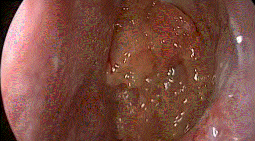
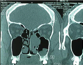
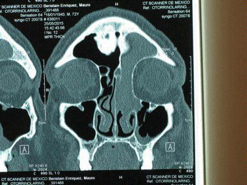
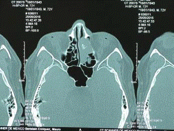
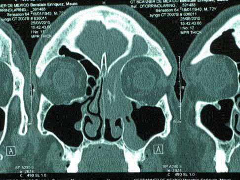
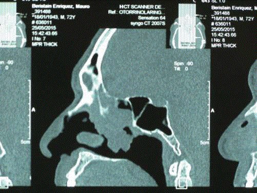
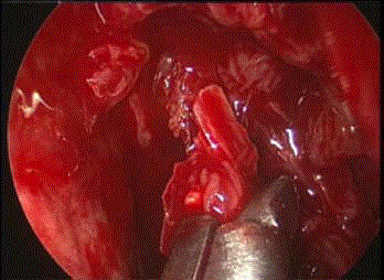
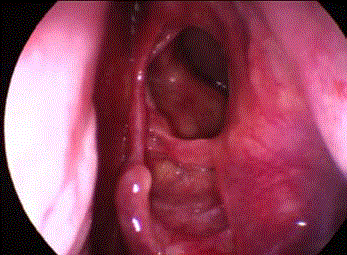
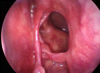
Male patient, 68 years-old that had been presenting clinical signs for 2 years, characterized by obstruction to the left nasal passage and occasional epistaxis. He goes to a medical practitioner who believes that the patient has a nasosinal polyp; the pathological result was that of inverted papilloma, for which he was referred to our otolaryngology department. The endoscopic image, typical of IP (Figure 1). The computerized tomography showed an opacity on the left nasal cavity, anterior ethmoidal cells and partially in the posterior ones; and on the frontal sinus (Figures 2 and 3). The oseitis zone on the anterior ethmoidal cells was identified (Figures 4-6). The endoscopic surgery was carried out using a microdebrider, with the aim of reducing the volume of the tumor and identifying the originating site (osteitis) (Figure 7). The tumor had lodged itself in the frontal sinus and had partially eroded the lamina papyracea. (Figures 8 and 9) are endoscopic images of the patient 3 years after his surgery and show no signs of recurrence.
Figure 1
Figure 2
Figure 3
Figure 3
The tumor extends to the ethmoid and frontal sinus. A magnetic
resonance image would be needed to differentiate tumor from mucus.
Figure 4
Figure 5
Figure 6
Figure 7
Figure 8
Figure 9
Discussion
If we strictly follow Dr. Krouse’s staging, this case would be a type 3 since it involved the frontal sinus. However, based on the theory of the originating site (osteitis), it is a type 2 and, statistically speaking, there is about a 3% chance of recurrence [8]. It has been 3 years since the patient underwent the surgery and most cases of recurrence happen within the first 2 years. Following protocol, all patients receive 5 years of follow-up. The classification should not be ignored, however, but rather be used to inform patients of the necessity of follow-up to detect possible recurrence.
Conclusion
The application of endoscopy for the resection of IP, along with the detection of osteitis as the tumor’s originating site, has led to a higher exit rate when treating IP, which is to say, less recurrence. The surgeon must be ready to perform radical surgery if the case calls for it, specifically if the originating site cannot be localized or if the tumor is multicentric.
References
- Roh HJ, Procop GW, Batra PS, Citardi MJ, Lanza DC. Inflammation and the pathogenesis of inverted papilloma. Am J Rhinol. 2004; 18(2):65-74.
- Chiu AG, Jackman AH, Antunes MB, Feldman MD, Palmer JN. Radiographic and histologic analysis of the bone underlying inverted papillomas. Laryngoscope. 2006; 116(9):1617-1620.
- Yousuf K, Wright ED. Site of attachment of inverted papilloma predicted by CT findings of osteitis. Am J Rhinol. 2007; 21(1):32-36.
- Lesperance MM, Esclamado RM. Squamous cell carcinoma arising in inverted papilloma. Laryngoscope. 1995; 105(2):178-183.
- Lawson, Ho BT, Shaari CM, Biller HF. Inverted Papilloma: A report of 112 Cases. Laryngoscope. 1995; 105(3):282-288.
- Krouse JH. Development of a staging system for inverted papilloma. Laryngoscope. 2000; 110(6):965-968.
- Healy DY, Chhabra N, Metson R, Holbrook EH, Gray ST. Surgical risk factors for recurrence of inverted papilloma. Laryngoscope. 2016; 126(4):796-801.
- Cannady SB, Batra PS, Sautter NB, Roh HJ, Citardi MJ. New staging system for sinonasal inverted papilloma in the endoscopic era. Laryngoscope. 2007; 117(7):1283-1287.

