Research Article
A Case of Furosemide Induced Nephrocalcinosis
Galesic Kresimir1, Tisljar Miroslav1*, Horvatic Ivica1, Stipcevic Mira2, Durlen Ivan1 and Galesic Ljubanovic Danica3
1Department of Nephrology, Dubrava University Hospital, Croatia
2Department of Cardiology, Dubrava University Hospital, Croatia
3Department of Pathology, Dubrava University Hospital, Croatia
*Corresponding author: Miroslav Tisljar, Department of Nephrology, Dubrava University Hospital, Zagreb, Croatia
Published: 14 Nov, 2017
Cite this article as: Kresimir G, Miroslav T, Ivica H, Mira
S, Ivan D, Danica GL. A Case of
Furosemide Induced Nephrocalcinosis.
Ann Clin Case Rep. 2017; 2: 1464.
Abstract
Background: Although furosemide is widely used for various medical conditions in adults, its
association with nephrocalcinosis is not well established. In adults, nephrocalcinosis induced
by furosemide is rare condition and presents as medullary nephrocalcinosis without significant
alteration of renal function.
Methods and Results: A 50-year-old female patient was admmited at our department due to renal
insufficiency (creatinine clearance was 62 mL/min) of unknown etiology. In medical history of the
patient, drug abuse was verified with a daily intake of 160 mg of furosemide during four years.
The reason of selfinitiated excessive furosemide intake was allegedly face and limb swelling. Patient
denied any previous kidney disease. The patient was normotensive with completely normal physical
status. Both, blood count and urinalysis were normal. Blood pH value was 7.43 and urine pH value
was 6.5. Amount of 24h urine proteinuria was 200 mg. Electrolites and urine acid concentrations
in blood and in 24h urine sample were normal. Serum levels of renin and aldosteron as well as
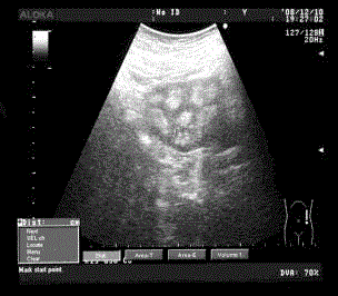
their ratio were regular. Urine concentration test showed izostenuric values. At sonography,
normal shaped and sized kidneys were found, with reduced parenchyma and a diffuse increase in
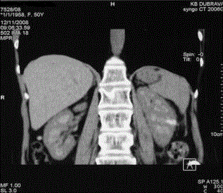
echogenicity throughout the medullary pyramids. By CT imaging, diffusely distributed soft tiny
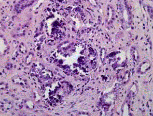
calcifications of kidneys medulla were identified. In kidney biopsy samples, medulla calcifications
(both in tubuli and interstitium) were described.
Conclusion: In conclusion, we can address that in our patient broad diagnostic procedure
was performed in a way we could exclude various causes of nephrocalcinosis. In adults with
nephrocalcinosis, besides many disorders, furosemide abuse should be considered as a potential
etiopatogenetic factor.
Keywords: Adults; Furosemide; Nephrocalcinosis; Renal insufficiency; Ultrasonography
Introduction
Nephrocalcinosis, the abnormal deposition of calcium salts in the renal parenchyma, is a rare
disease [1]. It can take two different forms: medullary nephrocalcinosis is the most frequent form
and is characterized by the exclusive involvement of the medullary pyramids. In a diffuse or cortical
nephrocalcinosis, which is rare, the entire parenchyma is affected: this pattern is associated with
severe metabolic disorders, such as hyperoxaluria, or end-stage nephropathy [2].
The association between nephrocalcinosis and long-term furosemide therapy is well documented
in infants [3]. Pyramidal involvement is present in more than 60% of preamature infants with low
birth weight or full term infants with congestive heart failure, treated with furosemide for long
periods. Although furosemide is widely used for various medical conditions in adults, its association
with nephrocalcinosis is not well established. In adults, nephrocalcinosis induced by furosemide is
rare and presents as medullary nephrocalcinosis without significant alteration of renal function. So
far, only a few cases of nephrocalcinosis caused by furosemide in adult patients have been described
in the literature, but just one of them included either imaging description of kidney and histological
documentation of the kidney biopsy [2].
A 50-year-old female patient was admitted to our hospital for investigation of azothaemia. She
was seen in another hospital for facial edema and swelling of the fingers and knees, and later was
referred to our hospital for further evaluation. In previous medical history, she had tonsillectomy,
fracture of left hand in a traffic accident. The patient entered menopause in her 37th year and was
taking oral hormonal supplementation only for several months. Three years before admission she had a parodontosis of upper teeth. During past several years she has
been occasionally suffering from insidious lumbar pain. The patient
has been self-administering furosemide 160mg/day for 4 years due
to persistent facial and knee edema and allegedly low daily urine
volume. Two weeks before hospitalization at our department she
had cancelled furosemide intake according to advice of her general
practitioner.
Methods and Results
On admission the patient was 161cm tall, weighted 53 kg and
had high blood pressure (170 mmHg/110 mmHg). No edema was
observed and physical examination was normal. We performed a
broad laboratory analysis. Both, blood count and urinalysis were
normal. Blood pH value was 7.43 and urine pH value was 6.5.
Urine microbiology was sterile. Amount of 24h urine proteinuria
(determined by Biuret test) was 200 mg. Initial metabolic evaluation
was performed: concentration of creatinine, calcium, phosphorus,
uric acid, sodium, potassium and chloride were measured both in
blood and urine. 24-urine samples were also analyzed for volume,
magnesium, oxalate and citrate.
Renal function was depressed (creatinine clearance was 62 ml/
min). All the other parameters analyzed were normal. A parathyroid
ultrasound was normal. Intact PTH was measured in the blood which
was normal. Serum levels of renin and aldosteron as well as their ratio
were regular. Urine concentration test showed izostenuric values (in
first urine sample osmolality was 290 mOsm/L). Finally, we decided
to make radio morfological and pathohistological investigation.
We used Simens acuson ultrasound device and Siemns Somatom
Sensation 16 CT system. A renal ultrasound showed medullary
nephrocalcinosis with total involvement of medullary pyramids
(Figure 1). An abnormal renal computerized tomography confirmed
this sonographic finding (Figure 2).
Biopsy findings
Three peaces of kidney tissue measuring 4-10 mm were analyzed.
About 70% of tissue was medulla and the rest was cortex with 11
glomeruli. Two glomeruli were globally sclerotic and the all others
had normal morphology. There were several foci of calcification in
tubules and interstitium predominantly within medulla (Figure 3).
Only few cortical tubules were involved by calcification. The all others
cortical tubules had normal morphology. The foci of calcification
were surrounded by interstitial fibrosis and tubular atrophy. The
blood vessels were unremarkable. There was no evidence of immune
deposits in immunofluorescent stains and electron microscopy
demonstrated the normal ultra structure of analyzed glomerulus and
surrounding tubules.
Clinical follow-up
Two months after discharged from hospital, patient came to
first control examination. In laboratory findings there were 24h
proteinuria level of 0.7 g and better renal excretion parameters: serum
creatinine 83 μmol/L with creatinine clearance of 72 mL/min and
normal serum potassium level (4.5 mmol/L).
Figure 1
Figure 2
Figure 3
Discussion
Nephrocalcinosis, is a term originally used to describe deposition
of calcium salts in the renal parenchyma due to hyperparathyroidism.
It is now more commonly used to describe diffuse, fine, renal
parenchymal calcification on radiology. Nephrocalcinosis can be
subdivided into the cortical type, which is classically the result of acute
tubular necrosis, and the medullary type seen in several metabolic
disorders. Many causes of nephrocalcinosis have been added since
the original description. These include causes of hypercalcemia and
hypercalciuria, medullary sponge kidney, hyperparathyreoidism,
renal tubular acidosis (RTA) specifically distal RTA, renal
tuberculosis, renal papillary necrosis, oxaluria, sarcoidosis and milkalkali
syndrome [4-7]. Furosemide is a powerful diuretic drug which
has been used successfully in the treatment of congestive heart failure.
Hyponatremia, hypokalemia and metabolic alkalosis are frequent
side-effects of the treatment. An association of renal calcinosis with
furosemide therapy in preterm infants was first reported in the
early 1980’s, the risk of developing nephrocalcinosis being highest in the most premature ones. Furthermore, the risk of developing
nephrocalcinosis was correlated with the dose of furosemide [2]. In
addition to, the period soon after initiation of furosemide therapy
appears most sensitive in regard of developing nephrocalcinosis [2].
The calciuric action of furosemide is one of the most distinguished
factors provoking nephrocalcinosis in preterm infants, but other
provoking pathogenic mechanisms are undoubtedly also involved in
most cases. Human and animal studies suggest that the development
of nephrocalcinosis does not always require hypercalciuria, but
alterations in urinary inhibitors of crystal formation (e.g. citrate,
magnesium), urinary excretion of oxalate and urine pH may also play
a role [1,8]. The possibility of nephrocalcinosis being due to type I
renal tubular acidosis must be excluded by a complete nephrologic
examination (serum potassium, pH, bicarbonate, tubular acidification
test with NH4Cl).
Our findings are consistent to previous reports suspecting longterm
furosemide treatment in adults may cause mild medullary
nephrocalcinosis, characterized by peripheral deposition of
calcium salts in the pyramids with depressed renal function. In
nephrocalcinosis, ultrasound examination shows an increase in
medullary echogenicitiy which can be massive or appear only as
an echogenic ring at the periphery of the renal pyramids [9]. This
pattern is an early manifestation of nephrocalcinosis [10]. This could
be useful information to sonographers involved in the diagnosis of
nephropathy. CT is the most accurate and sensitive technique and
therefore the modality of choice [2]. In conclusion, we considered
that furosemide treatment should be part of differential diagnosis
list of medullary nephrocalcinosis in adults undergoing long-term
therapy with this drug, especially if the high dose of drug is used as
in this case.
References
- Alon US. Nephrocalcinosis. Curr Opin Pediatr. 1997; 9: 160-165.
- Kim YG, Kim B, Kim MK, Chung SJ, Han HJ, Ryu JA, et al. Medullary nephrocalcinosis associated with long-term furosemide abuse in adults. Nephrol Dial Transplant. 2001; 16: 2303-2309.
- Hufnagle KG, Khan SN, Penn D, Cacciarelli A, Williams P. Renal calcifications: a complication of long-term furosemide therapy in preterm infants. Pediatrics. 1982; 70:360-363.
- Jacobs TP, Bilezikian JP. Clinical review: Rare causes of hypercalcemia. J Clin Endocrinol Metab. 2005; 90: 6316-6322.
- McGuinness B, Logan JI. Milk alkali syndrome. Ulster Med J. 2002; 71: 132-135.
- Garel L, Filiatrault D, Robitaille P. Nephrocalcinosis in Bartter's syndrome. Pediatr Nephrol. 1988; 2: 315-317.
- Herlitz LC, Bruno R, Radhakrishnan J, Markowitz GS. A case of nephrocalcinosis. Kidney Int. 2009; 75: 856-859.
- Wardener HE. Idiopathic edema: role of diuretic abuse. Kidney Int. 1981; 19: 881-891.
- Simoes A, Domingos F, Prata MM. Nephrocalcinosis induced by furosemide in an adult patient with incomplete renal tubular acidosis. Nephrol Dial Transplant. 2001; 16: 1073-10744.
- al-Murrani B, Cosgrove DO, Svensson WE, Blaszczyk M. Echogenic rings--an ultrasound sign of early nephrocalcinosis. Clin Radiol. 1991; 44: 49-51.


