Clinical Image
Emphysematous Pyelonephritis
Ana Ruiz*, Clément Fabre, Jean Rémy Boutault and Catherine Merzeau
Department of Radiology, Hôpital Gaston Bourret, Nouvelle Calédonie
*Corresponding author: Ana Ruiz, Service de Radiologie, Hôpital Gaston Bourret, 7 avenue Paul Doumer, BP J5, 98849 Nouméa Cedex, Nouvelle Calédonie
Published: 10 Nov, 2017
Cite this article as: Ruiz A, Fabre C, Boutault JR, Merzeau
C. Emphysematous Pyelonephritis. Ann
Clin Case Rep. 2017; 2: 1460.
Abstract
In this article, typical images of emphysematous pyelonephritis are displayed through a clinical case. This diagnosis must be quickly recognized to achieve appropriate medical and surgical treatment.
Keywords: Emphysematous pyelonephritis; Computed tomography; Nephrectomy
Case Presentation
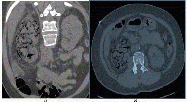
A 45-year-old woman with no known medical history was referred to our institution for abdominal pain, diarrhea, vomiting, and fever. Laboratory findings showed inflammation (C-reactive protein: 356 mg/L), renal failure (serum creatinine: 156 μmol/L), thrombocytopenia (platelet : 88x109/L) and hyperglycemia (Serum Glucose: 28.3 mmol/L). Urinary test strips showed leukocyturia and hematuria [1-3]. An unenhanced computed tomography scan of the abdomen demonstrated the presence of gas in all the right renal parenchyma, the collecting system and the perinephric tissue, concluding in a class 3A emphysematous pyelonephritis, without urinary tract obstruction (Figure 1a and b). The patient was treated with broad-spectrum intravenous antibiotics. Clinical and biological worsening conducted to surgical treatment. Right nephrectomy was performed and purulent liquefaction of the right kidney was observed. After the surgery, the patient improved significantly. The urine cultures showed significant growth of Escherichia coli (E. coli).
Figure 1
Figure 1
a) and b) An unenhanced computed tomography scan of the abdomen demonstrated the presence of
gas in all the right renal parenchyma, the collecting system and the perinephric tissue, concluding in a class 3A
emphysematous pyelonephritis, without urinary tract obstruction.
References
- Huang JJ, Tseng CC. Emphysematous pyelonephritis: clinicoradiological classification, management, prognosis, and pathogenesis. Arch Intern Med. 2000; 160:797-805.
- Grayson DE, Abbott RM, Levy AD, Sherman PM. Emphysematous infections of the abdomen and pelvis: a pictorial review. Radiographics. 2002; 22: 543-61.
- Somani BK, Nabi G, Thorpe P, Hussey J, Cook J, N'Dow J, et al. Is percutaneous drainage the new gold standard in the management of emphysematous pyelonephritis? Evidence from a systematic review. J Urol. 2008; 179: 1844-9.

