Case Report
Surgical Treatment for Chronic-Contained Rupture of Thoracic Aortic Aneurysm Complicated with Vertebral Erosion: Case Report
Alexey Nesmachnyy*, Alexander Chernyavskiy, Julia Kareva, Dmitriy Sirota and Alexandra
Tarkova
Department of the Aorta and Coronary Arteries Surgery, Siberian Biomedical Research Center Ministry of Health
Russian Federation, Russia
*Corresponding author: Alexey Nesmachnyy, Department of the Aorta and Coronary Arteries Surgery, Siberian Biomedical Research Center Ministry of Health Russian Federation, 15 Rechkunovskaya St., Novosibirskaya Region, Novosibirsk, Russia
Published: 28 Aug, 2017
Cite this article as: Nesmachnyy A, Chernyavskiy A,
Kareva J, Sirota D, Tarkova A. Surgical
Treatment for Chronic-Contained
Rupture of Thoracic Aortic Aneurysm
Complicated with Vertebral Erosion:
Case Report. Ann Clin Case Rep. 2017;
2: 1425.
Abstract
In this article, we present the clinical case and successful surgical treatment of aneurysm of the arch and the descending aorta with the destruction of the vertebral bodies.
Keywords: Aortic aneurysm; Thoracic; Aortic rupture; Thoracic vertebrae; Spinal diseases
Introduction
Recent clinical literature has described only few cases of erosion of the spine caused by a chronic abdominal aortic aneurysm [1-3]. Aneurysm of the arch and the descending aorta along with vertebral destruction is a very rare combination with no observations in the global clinical practice. Only one scientific review has mentioned a similar pathology [4].
Case Presentation
A 43-year-old man was admitted to the hospital with complaints of foreign body sensation,
periodic pressing chest pain, shortness of breath on slight exertion, and paroxysmal dry cough.
According to anamnesis, at the age of 20, the patient had a fall from approximately 6-m height.
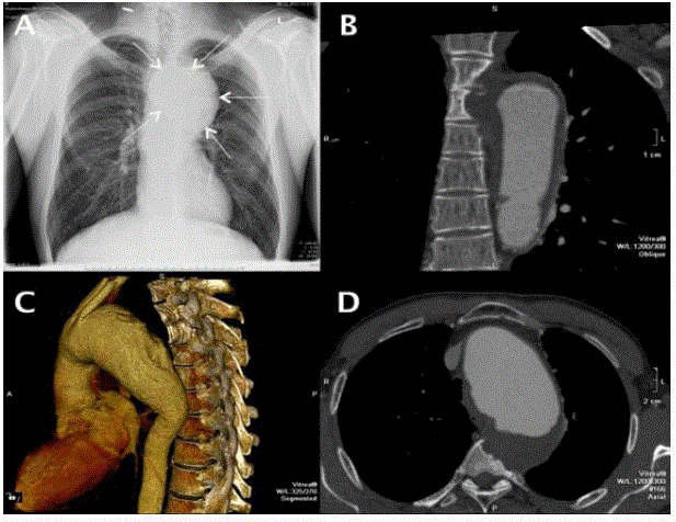
His six-month-old radiograph showed an aneurysm of the thoracic aorta (Figure 1a). Multi-Spiral
Computed Tomography (MSCT) angiography identified signs of aneurysm of the thoracic aorta
that infiltrated its wall, neoplasm of the upper floor of the posterior mediastinum, and destruction
of vertebral bodies (Th3, Th4, And Th5) and fourth rib on the left (Figure 1b-d). Considering failure of the aortic arch with the spread of the descending thoracic aorta, it was clear that stenting of
this segment should be performed after the preparatory stage of the brachiocephalic arterial switch operation. In addition, given the probability of finding a tumor mass
in the projection of the aneurysm, an open heart surgery was decided
to be performed.
Operative technique
The operation was planned as follows: after the median
sternotomy, cardiopulmonary bypass connection was provided;
arterial cannula was inserted into the right subclavian artery, and
venous cannula into the right atrium; cardiopulmonary bypass was
initiated; and the patient’s body was cooled to 24°C.
On visualization of the aorta, its diameter at the sinotubular
junction was about 30-35 mm and that at the level of the ascending
portion and the aortic arch was 60 mm. Major inflammation was noted
in the projection of the upper and middle third of the descending
aorta, distal to the arch; the aorta soldered to the upper lobe of
left lung and vertebral bodies, th3 and th4. When the temperature
reached 24°C, cardiopulmonary bypass was abandoned; this was
followed by antegrade perfusion to the brain at a flow rate of 15 ml/
kg/minute, and aortotomy was performed. The lumen of the aorta had
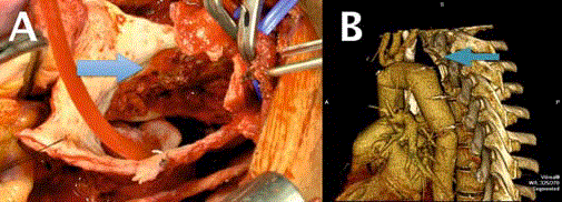
thrombotic masses with varying degrees of organization. Vertebral
bodies with erosive changes were visualized on these masses after
they were removed (Figure 2a). According to express histological
analysis, a modified aneurysmal aortic wall tissue was involved
in the destruction of the vertebral bodies. Sinuses of valsalva were
not expanded, when viewed from the tricuspid aortic valve without
pathology. Prosthesis in the ascending portion of the arch and the
descending thoracic aorta at the level of the 5-6, and brachiocephalic
arteries were manipulated using vascular prosthesis (vascutek siena
plexus 26). On the fourth day after surgery, the patient was transferred
from the intensive care unit to the profile department. During postoperative
period, the patient showed symptoms of respiratory failure,
severe asthenic syndrome, signs of exudative pleurisy that required
pleural puncture, and prolonged antibiotic therapy (sulperazon) due
to the long sub-febrile fever. On the forty-sixth day after operation,
the patient left the hospital in good condition. Post-operative most
showed defects on the left surface of the vertebrae th3-th4-th5 and
the head of the fourth rib at the level of diligence modified paraaortic
tissue. This is a type of atrophy caused by chronic aneurysmal
pressure (Figure 2b).
Figure 1
Figure 1
(A) Images of X-ray of the chest. The arrow indicates the aneurysm of the thoracic aorta. (B-D) Images
of multi-spiral CT angiography of the chest before surgery.
Figure 2
Figure 2
(A) Intra-Operative Photo. The Arrow Indicates the Site of the
Destruction of the Vertebral Bodies Th3, Th4. (B) Image of the Post-Operative
Multi-Spiral CT Angiography of the Chest. The Arrow Indicates Defects On
The Left Surface Of The Vertebrae Th3-Th4-Th5.
Discussion
Chronic-Contained Rupture of the aorta is relatively rare in the
abdomen (2.7% of cases have reported pathology of the abdominal
aorta) and very rare in the thoracic aorta [1]. In the literature, only
few similar clinical cases have been described [4]. Patients with this
condition are usually haemodynamically stable and do not perform
acute symptoms, although they always have a high risk of rupture
of the aortic wall. Persistent pressure on the surrounding tissue
due to pulsation of the aorta over a period is probably the cause of
destruction of solid structures such as vertebral bodies. In such
cases, a differential diagnosis must be made using msct between
primary tumours or metastatic vertebral tumours, vertebral fractures,
osteoporosis, and other possible diseases [3,5].
Concerning Surgical Treatment, aggressive removal of
aneurysmal wall near the spinal cord is not recommended because
of possible damage to the spinal cord. Sometimes, it is sufficient
to eliminate the pressure pulsating aneurysmal wall to prevent
compression of the surrounding tissues. In such cases, it is possible
to insert a stent-graft in the absence of inflammation. However, there
are reports that indicate the progress of symptoms of compression in
the surrounding tissues after the stent-graft, thus requiring another
open intervention in the long term.
In the clinical case presented here, the strategy of performing an
open heart surgery was chosen as a tumour process was suspected in
the projection of the aneurysm, and the spinal cord was located at a
sufficient distance from the site of destruction of the vertebrae (as
seen in the most findings).
References
- Lai CC, Tan CK, Chu TW, Ding LW. Chronic Contained Rupture Of An Abdominal Aortic Aneurysm With Vertebral Erosion. Can Med Assoc J. 2008; 178: 995-996.
- Arici V, Rossi M, Bozzani A, Moia A, Odero A. Massive Vertebral Destruction Associated With Chronic Rupture Of Infrarenal Aortic Aneurysm: Case Report And Systematic Review Of The Literature In The English Language. Spine (Phila Pa 1976). 2012; 37: 1665-1671.
- Jang JH, Kim HS, Kim SW. Severe Vertebral Erosion by Huge Symptomatic Pulsating Aortic Aneurysm. J Korean Neurosurg Soc. 2008; 43: 117-118.
- Yuksekkaya R, Koner AE, Celikyay F, Beyhan M, Almus F, Acu B. Multidetector Computed Tomography Angiography Findings Of Chronic Contained Thoracoabdominal Aortic Aneurysm Rupture With Severe Thoracal Vertebral Body Erosion. Case Rep Radiol 2013; 2013: 1-4.
- Matsunaga I, Nogami E, Higuchi S, Okazaki Y, Itou T. Chronic Contained Rupture of Aortic Aneurysm with Thoracic Vertebral Erosion. Asian Cardiovasc Thorac Ann. 2015; 23: 564-566.


