Case Report
Extrusion of Ventricular-Peritoneal Shunt through the Urethra: Pediatric Case Report and Literature Review
Ailsa May Li Gan, Linnea D Duke, Robert Schwarz and Nicholas Power*
Department of Urology, Victoria Hospital, London
*Corresponding author: Nicholas Power, Department of Urology, London Health Sciences Centre, Victoria Hospital 800 Commissioners Road East London, ON, Canada N6A 5W9, UK
Published: 21 Aug, 2017
Cite this article as: Gan AML, Duke LD, Schwarz R, Power
N. Extrusion of Ventricular-Peritoneal
Shunt through the Urethra: Pediatric
Case Report and Literature Review.
Ann Clin Case Rep. 2017; 2: 1423.
Abstract
Ventricular-peritoneal shunts are commonly used to treat hydrocephalus, but carry the risk of complications including perforation of abdominal viscera. Bladder perforation by the distal shunt may present with urinary symptoms uncommonly seen in children, raising the clinical suspicion of this rare phenomenon. Even less commonly, this complication can result in extrusion of the distal catheter through the urethra, which has only previously been reported four times in males. In this paper, we discuss the existing literature and present the youngest male case (2 years old) of ventricular-peritoneal shunt bladder perforation with urethral extrusion. Following, we review the existing literature using a systematic search of PUBMED and MEDLINE.
Keywords: Ventricular-peritoneal shunt (V-P); Cerebral spinal fluid (CSF) shunt; Shunt migration; Foreign body migration; Shunt complications
Introduction
The surgical insertion of a ventricular-peritoneal (V-P) shunt is the most common method of treatment for hydrocephalus [1]. V-P shunts allow cerebrospinal fluid to drain via a catheter into the peritoneal cavity. The most commonly reported complications of V-P shunts include shunt obstruction and infections such as intra-abdominal abscess or bacterial meningitis [2]. The percentage of abdominal complications associated with V-P shunts has been reported to range from 5% to 47%, and include formation of peritoneal pseudocysts, formation of incisional hernias, and subcutaneous collection of cerebrospinal fluid (CSF) [1]. Migration of the distal catheter infrequently results in perforation of both solid and hollow viscera. Extrusion of catheters through the mouth, anus, umbilicus, scrotum, and vagina has been reported in the literature [3-8]. However, migration of the distal V-P shunt and perforation of the bladder, in particular, is uncommon. Anatomically, the bladder poses a difficult target for the distal shunt, as the catheter would have to either pass through the peritoneum and into the extra-peritoneal space before reaching the wall of the bladder, or penetrate the peritoneal lining directly adjacent to the bladder dome thus entering the bladder lumen [9]. The case presented in this paper is the youngest male case of bladder perforation presenting with urethral extrusion. This represents an uncomplicated case occurring in a patient with a normal bladder.
Case Presentation
We report a two-year-old boy with a history of surgical resection of a posterior fossa
meduloblastoma and left V-P shunt placement in 2008. Eight months after placement of the V-P
shunt, the boy presented to the IWK Hospital in Halifax, Nova Scotia, after his mother discovered
a tube extruding from his urethra, concerned that the infant had inserted the tube himself. Closer
examination at the hospital revealed it was, in fact, the peritoneal portion of his V-P shunt extruding
through his urethra (Figure 1).
The patient’s vital signs were stable. Physical examination revealed no other findings. Urinalysis
was normal. A radiographic shunt study including X-ray of the skull, chest, and abdomen revealed
the V-P shunt to be continuous and followed the route of the left pelvic ureter (Figure 2). Perforation of the bladder was noted. No evidence of bowel obstruction, free air, or fluid was found. Renal
ultrasound revealed no evidence of perforation of the kidneys by the V-P shunt. No evidence of
hydronephrosis, hydroureter, urinary ascites, perirenal, or perivesicle fluid collection was observed.
However, ultrasound of the bladder revealed the presence of a tubular structure within its lumen
(Figure 3). At the time of presentation, the V-P shunt was believed to be non-functional and was surgically removed without complication on the same day. A Foley catheter was inserted at the time of shunt removal to prevent bladder distension and to allow
healing at the site of bladder wall perforation. The Foley catheter was
removed on post-operative day five after which the patient was able to
void normally on his own. The patient was discharged from hospital
in stable condition on post-operative day six.
Methods
A literature search was performed using MEDLINE and PubMed using the keywords “ventricular-peritoneal shunt”, “ventriculoperitoneal shunt”, “cerebrospinal fluid shunt”, “bladder perforation”, and “migration”. A second manual search through references from selected articles provided additional sources, completing the search.
Figure 1
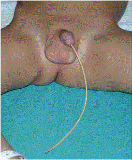
Figure 1
Distal aspect of the ventriculoperitoneal shunt protruding via the
urethra.
Figure 2
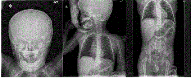
Figure 2
Shunt series tracing the course of the ventriculoperitoneal shunt
catheter.
Figure 3
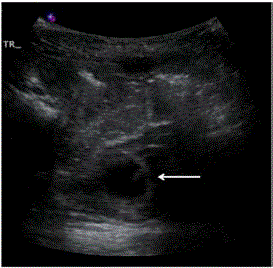
Figure 3
Ultrasound image of the bladder demonstrating the presence of a
tubular structure (indicated by arrow) within its lumen.
Discussion
We found a total of 25 cases of V-P shunt with bladder perforation
reported in 21 different articles in the literature [9-25]. Of the 25 cases,
22 reported the age of the patient, which ranged from 8 days [26] to
82 years [22], with the majority (19/22) occurring in children aged 16
or younger. This may be primarily due to the fact that V-P shunts are
more prevalent in this age group as hydrocephalus commonly occurs
with congenital spinal and neural tube defects [27]. Furthermore,
these defects are often associated with neurogenic bladder, requiring
augmentation. Bladder perforation by V-P shunt can occur in both
augmented bladders and normal bladders.
Through our literature search, we found only twelve cases
described to present with urethral extrusion of the distal catheter
[9,12,13,16-19,21-23,26,27]. From these cases, urethral extrusion
appears to preferentially effect females, presenting at a male to female
ratio of 1:2. It is possible that this is due to the anatomical differences between sexes, predisposing females with shorter, straighter urethras to extrusion.
Cases without extrusion presented clinically with urinary
symptoms including at least one of dysuria, urgency, retention,
and lower abdominal pain. Acute bladder perforation is likely to
present with a degree of hematuria, but interestingly, the present case
demonstrated normal urinalysis. It is possible that the perforation of
the bladder occurred sometime before extrusion, allowing adequate
healing to occur before presentation to the emergency department
upon extrusion of the distal catheter.
The present case represents an occurrence of a bladder perforation
by distal V-P shunt with no additional pathology. However, bladder
perforation can present concurrently with local complications
such as bladder calculi [2,11,20], or even meningitis as a result of
bacterial ascent to the proximal V-P shunt [26]. Rarely, it has been
documented to occur with knotting of the distal shunt, requiring
a more complicated procedure of removal due to the inability to
remove the shunt atraumatically from the urethra [9,16]. Knotting of
the catheter has been hypothesized to be due to retained memory by
the shunt material of the packaged shape [28].
Migration of the distal V-P shunt is not well understood, but has
been suggested to be the result of inflammatory reactions within the
peritoneal cavity due to interactions between the shunt apparatus,
the CSF contents, and the peritoneal membrane as a foreign body
response is initiated [29]. In addition, bowel peristalsis, increased
intra-abdominal pressure from inspiration, expiration, and valsalva
effects, and CSF pulsations may also factor into friction effects that
lead to visceral involvement [16,23]. The resultant inflammatory
reactions can then lead to fixation of the distal V-P shunt, progressing
to erosion of the visceral or abdominal wall, finally resulting in
complications such as perforation, local abscess formation, and
shunt obstruction [26]. Prasad et al. described the first reported
case of urethral extrusion following bladder perforation of the distal
V-P shunt, postulating a mechanism for this complication. They
propose four events to occur: fixation (as described earlier to involve
inflammatory reactions), penetration, perforation, entry into the
urethra, and finally extrusion [19]. It is unclear how long this process
takes in vivo. There has been a variable amount of time reported
from the initial placement of the shunt to the presentation of bladder
involvement ranging from three months to twelve years, with the
exception of one case that presented after one day [26]. However, the
latter case is more likely to have been caused iatrogenically at surgical
insertion with a trocar. It is likely that perforation of the bladder can occur and remain asymptomatic for some time before presenting to clinic.
In terms of prevention, it has been recommended that the length
of the V-P shunt be taken into consideration and controlled to
reduce the degree of unnecessary rubbing that may be contributing
to migration complications [2,12]. In addition, the material of the
shunt should be considered, as the previously mentioned memoryretaining
properties of the shunt may increase the risk of coiling,
knotting, and friction in general within the peritoneal cavity.
Finally, careful surgical placement of the shunt if a trocar is used is
imperative to avoid direct perforation of any visceral organs leading
to acute complications. Having patients empty their bladder prior to
the procedure, particularly toddlers with smaller intra-abdominal
compartments, is recommended. Fortunately, the present case had
a positive outcome with no further complaints related to the bladder
perforation and extrusion of the shunt. This is in agreement with
the general outcome reported in the literature. However, due to the
fact that this complication requires surgical management and can
progress to serious additional shunt-related morbidity, it is important
for physicians treating patients with V-P shunts to be aware of this
rare complication and monitor for urinary symptoms that may
represent early indicators of urogenital involvement.
References
- Nfonsam V, Chand B, Rosenblatt S, Turner R, Luciano M. Laparoscopic management of distal ventriculoperitoneal shunt complications. Surg Endosc Other Interv Tech. 2008; 22: 1866-1870.
- Xu S, Sheng W, Qiu Y, Wang J. An unusual complication of ventriculoperitoneal shunt: urinary bladder stone case report and literature review. Iran Red Crescent Med J. 2016; 18: e26049.
- Yilmaz MB, Egemen E, Tonge M, Kaymaz M. Transoral protrusion of a peritoneal catheter due to gastric perforation 10 years after a ventriculoperitoneal shunting: Case report and review of the literature. Turk Neurosurg. 2013; 23: 285-288.
- Esposito C, Porreca A, Gangemi M, Garipoli V, De Pasquale M. The use of laparoscopy in the diagnosis and treatment of abdominal complications of ventriculo-peritoneal shunts in children. Pediatr Surg Int. 1998; 13: 352-354.
- Akcora B, Serarslan Y, Sangun O. Bowel perforation and transanal protrusion of a ventriculoperitoneal shunt catheter. Pediatr Neurosurg. 2006; 42: 129-131.
- Walsh AR, Kombogiorgas D. Coiled ventricular-peritoneal shunt within the scrotum. Pediatr Neurosurg. 2004; 40: 257-258.
- Wani AA, Ramzan A, Wani MA. Protrusion of a peritoneal catheter through the umblicus: An unusual complication of a ventriculoperitoneal shunt. Pediatr Surg Int. 2002; 18: 171-172.
- Nagulic M, Djordjevic M, Samardzic M. Peritoneo-vulvar catheter extrusion after shunt operation. Child’s Nerv Syst. 1996; 12: 222-223.
- Pohlman GD, Wilcox DT, Hankinson TC. Erosive bladder perforation as a complication of ventriculoperitoneal shunt with extrusion from the urethral meatus: Case report and literature review. Pediatr Neurosurg. 2012; 47: 223-226.
- Binning MJ, Ragel BT, Walker ML, Kestle JR. Retained peritoneal shunt tubing causing hematuria. Case illustration. J Neurosurg. 2006; 104: 434.
- Butler L, Keys C, Lam JPH. Bladder calculus formation on the tip of a migrated disused ventriculoperitoneal shunt. J Pediatric Surg. 2013; 48: e1-e3.
- Chen TH, Lin MS, Kung WM, Hung KS, Chiang YH, Chen CH. Combined ventriculoperitoneal shunt blockage, viscus perforation and migration into urethra, presenting with repeated urinary tract infection. Ann R Coll Surg Engl. 2011; 93: 151-153.
- De Aguiar GB, Mizrahi C, Aquino JHW, Tavares CM, Telles C, Nigri F, et al. Urethral extrusion of a peritoneal catheter in a patient with a neobladder: A rare complication of shunt insertion. Neuropediatrics. 2011; 42: 124-127.
- De Jong L, Van Der Aa F, De Ridder D, Van Calenbergh F. Extrusion of a ventriculoperitoneal shunt catheter through an appendicovesicostomy. Br J Neurosurg. 2011; 25: 115-116.
- Eichel L, Allende R, Mevorach RA, Hulbert WC, Rabinowitz R. Bladder calculus formation and urinary retention secondary to perforation of a normal bladder by a ventriculoperitoneal shunt. Urology. 2002; 60: 344.
- Kataria R, Sinha VD, Chopra S, Gupta A, Vyas N. Urinary bladder perforation, intra-corporeal knotting, and per-urethral extrusion of ventriculoperitoneal shunt in a single patient: Case report and review of literature. Child’s Nerv Syst. 2013; 29: 693-697.
- Mevorach RA, Hulbert WC, Merguerian PA, Rabinowitz R. Perforation and intravesical erosion of a ventriculoperitoneal shunt in a child with an augmentation cystoplasty. J Urol. 1992; 147: 433-434.
- Mihajlovic M, Tasic G, Raicevic M, Mrdak M, Petrovic B, Radlovic V. Asymptomatic perforation of large bowel and urinary bladder as a complication of ventriculoperitoneal shunt: report of two cases. Srp Arh Celok Lek. 2012; 140: 211-215.
- Prasad VS, Krishna AM, Gupta PK. Extrusion of peritoneal catheter of ventriculoperitoneal shunt through the urethra. Br J Neurosurg. 1995; 9: 209-210.
- Ramana Murthy KV, Jayaram Reddy S, Prasad DVSRK. Perforation of the distal end of the ventriculoperitoneal shunt into the bladder with calculus formation. Pediatr Neurosurg. 2009; 45: 53-55.
- Surchev J, Georgiev K, Enchev Y, Avramov R. Extremely rare complications in cerebrospinal fluid shunt operations. J Neurosurg Sci. 2002; 46: 100-102.
- Ueda Y, Kakino S, Hashimoto O, Imoto K. Perforation of the bladder by a peritoneal catheter: An unusual late complication of ventriculo-peritoneal shunt. Neurol Surg. 1998; 26: 413-416.
- Yazar U, Kanat A, Akca N, Gazioglu G, Arda IS, Kazdal H. Urethral protrusion of the abdominal catheter of ventriculoperitoneal shunt: Case report of extremely rare complication. J Pediatr Neurosci. 2012; 7: 111-113.
- Yerkes EB, Rink RC, Cain MP, Luerssen TG, Casale AJ. Shunt infection and malfunction after augmentation cystoplasty. J Urol. 2001; 165: 2262-2264.
- Grosfeld JL, Cooney DR, Smith J, Campbell R. Intra-abdominal complications following ventriculoperitoneal shunt procedures. Pediatrics. 1974; 54: 791-796.
- Mutlu M, Kader S, Aslan Y, Yazar U, Imamoglu M. An acute complication of ventriculoperitoneal shunt with bladder perforation and extrusion through the urethra in a newborn: Case report and review of the literature. Pediatr Neurosurg. 2015; 50: 264-269.
- Miranda ME, de Sousa MB, Tatsuo ES, Quites LV, Giannetti AV. Bladder perforation by ventriculoperitoneal shunt. Child’s Nerv Syst. 2016; 32: 2321-2326.
- Dominguez CJ, Tyagi A, Hall G, Timothy J, Chumas PD. Sub-galeal coiling of the proximal and distal components of a ventriculo-peritoneal shunt. An unusual complication and proposed mechanism. Childs Nerv Syst. 2000; 16: 493-495.
- Aquino HB, Carelli EF, Borges Neto AG, Pereira CU. Nonfunctional abdominal complications of the distal catheter on the treatment of hydrocephalus: An inflammatory hypothesis? Experience with six cases. Child’s Nerv Syst. 2006; 22: 1225-1230.