Case Report
An Omental Mass: Any Idea?
Mascianà G*, Gallo IF, Valeri S and Coppola R
Department of General Surgery, University Campus Bio-Medico, Italy
*Corresponding author: Mascianà Gianluca, Università Campus Bio-Medico, Via Alvaro del Portillo 21 - 00128 Rome, Italy
Published: 20 Jul, 2017
Cite this article as: Mascianà G, Gallo IF, Valeri S, Coppola
R. An Omental Mass: Any Idea?. Ann
Clin Case Rep. 2017; 2: 1406.
Abstract
Pseudomyxoma Extra Peritonei (PE) is a rare finding, the most common cause is the rupture of a mucocele of the appendix into the retroperitoneum. Here we report a case of a 52 years old female patient with a mass in the right abdomen and vague lower abdominal pain underwent resection of an extraperitoneal encapsulated mass. The histopathological examination revealed a mucinous pseudomyxoma with a low grade of differentiation. We report a case of pseudomyxoma extra peritonei with a review of literature.
Case Presentation
A 52-year-old female patient was referred to our hospital for a mass in the right abdomen and
vague lower abdominal pain. The only remarkable event in her past history was a right breast fibroid
neoplasm that had been removed 10 years before.
Investigations
An abdominal examination revealed a large, fixed mass in the subcostal right region. There were
no cervical, axillary or inguinal lymphadenopathies, nor any signs of ascites. Other tests were normal
with the exception of a slight increase in the CEA level (8.5 ng/ml; normal value: 0-2.5). Computed
tomography (CT) of the chest and abdomen revealed a roundish, 100 mm x 70 mm x 80 mm mass in
the right flank, with regular margins, a partial capsule and a19-mm internal calcification with mural
calcification and hyperdense central striae (Figure 1). The US-guided aspiration of the mass yielded a cellular material. The upper and lower endoscopic examinations were negative.
Treatment
The case was discussed ata multidisciplinary team meeting. As it proved difficult to make
a preoperative diagnosis, the patient underwent surgery, during which an extraperitoneal
encapsulated mass that was free of all surrounding structures, including the pancreas, kidneys
and bowel, was found. This mass was resected completely without breaking the capsule. The
histopathological analysis revealed a mucinous pseudomyxoma with a low grade of differentiation.
Immunohistochemistry was positive for CK20 and CK7. The patient was discharged 4 days after
surgery without any complications. At follow-up, 3 years later, there was no sign of recurrence.
Figure 1
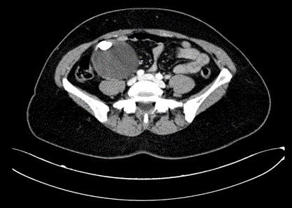
Figure 1
CT scan of the abdomen and pelvis, demonstrating the mass in the right abdomen.
Table 1

Table 1
37 reported cases appendix.
Figure 2
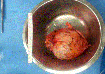
Figure 2
Surgical specimen.
Discussion
Werth [1] first described pseudomyxoma peritonei in 1884 as the presence of mucinous and gelatinous material in the peritoneal cavity. In 1948, Bonann [2] reported a pseudomyxoma involving the retroperitoneum alone, which was subsequently displayedby Coppini [3] in 1950. Twenty years later, Early [4] described a retroperitoneal mucocele of the appendix that contained 10 litres of mucus that had not ruptured, thus allowing a complete curative excision to be performed; this was referred to by Moran [5] as pseudomyxoma extra peritonei (PE) in 1988. Shelton et al. [6] later named it Pseudomyxoma retroperitonei. Pseudomyxoma retroperitoneal is a rare disease of which there are only 37 reported cases in the literature; the most common cause is the rupture of a mucocele of the appendix into the retroperitoneum (Table 1). It affects both sexes to the same extent, prevalently after the age of 60 years [7], with other potential primary sites including a mucinous neoplasm of the ovary or bowel, or a primary retroperitoneal mucinous cystadenoma/cystadenocarcinoma; a histopathological analysis reveals aggregates of mucus and epithelial cells displaying varying degrees of atypia and differentiation. The cells are generally positive for CK20 and negative for CK7 [8]. Pseudomyxoma can be classified as grade I or benign disseminated peritoneal adenomucinosis, as grade II or intermediate subtype, and as grade III or malignant peritoneal mucinous carcinomatosis [9,10]. In the majority of cases the pathogenesis of Pseudomyxoma retroperitonei is explained by a leak through the peritoneum (retroperitoneal presentation associated with intraperitoneal pseudomyxoma). In the absence of peritoneal pseudomyxoma, a variant of the anatomy of the appendix (retroperitoneal location) may explain the extraperitoneal pseudomyxoma, though this hypothesis is still speculative [11]. A preoperative diagnosis is very rare; symptoms such as fatigue, decreased appetite with weight loss, the presence of a palpable mass and slowly progressing abdominal or lumbar pain are common. CEA and CA 19.9 are reported to be increased in 56% - 75% and 58% - 67% of patients, respectively [10]. Ultrasound may detect the mucina as retroperitoneal fluid and help to make a diagnosis by means of needle aspiration, while CT with intravenous, oral and rectal contrast may distinguish the mucinous substance from the normal watery fluid by means of density property analysis (5-20 Hounsfield units for mucous vs 0 Hounsfield units for water) [9]. At CT, Pseudomyxoma retroperitonei appears as a mass that is often multicystic, has septa or thick walls and may be characterized by mural calcifications that displace adjacent structures [12]. Surgery The treatment of pseudomyxoma differs substantially depending on whether it is intraperitoneal or extraperitoneal. For intraperitoneal pseudomyxoma, Sugarbaker et al. [13] recommended an aggressive, complex surgical procedure that involves extirpation of the mucinous material, debulking and peritonectomy in order to remove as much macroscopic disease as possible (cytoreductive surgery, CRS) using heated intraperitoneal hyperthermic chemotherapy (HIPEC). By contrast, the recommended treatment for extraperitoneal pseudomyxoma is, as for benign disease, resection of the site of origin sometimes followed by systematic chemotherapy [7]. Glehen et al. [14] reported a median survival of 156 months, with 5- and 10-year survival rates of 72% and 55%, respectively, in 501 pseudomyxoma peritonei patients who had undergone CRS (complete or incomplete) followed by HIPEC. The majority of the patients (~70%) underwent complete cytoreduction. This uniform treatment approach has led to a better 10-year survival than that recorded in historical controls [15,16]. Although no data are available on the use of hyperthermic retroperitoneal chemotherapy, this treatment should be considered owing to the high recurrence rate [7]. Surgery The risk of recurrence is such that follow-up, based on a physical examination, CT scan and serum markers, is essential. Combined treatment in Pseudomyxoma retroperitonei is associated with a 20- year survival rate in up to 70% of patients [17], whereas the survival rate for pseudomyxoma intra-peritonei, in which vital abdominal structures are involved, is shorter [5].
References
- Werth R. Pseudomyxomaperitonei. Arch Gynaecol. 1884; 24: 100–118.
- Bonnan LJ, Davis JG. Retroperitoneal mucocoele of the appendix. A case report with characteristic roentgen features. Radiology. 1948; 51: 375–382.
- Coppini B. Su di un rarissimo caso di pseudomixoma retroperitoneale di probabile origine appendicolare. G Clin Med. 1950; 31: 601–612.
- Early KS, Stephenson DV, Davis WC. Giant retroperitoneal mucocele simulating pseudomyxomaperitonei and mucinous adenocarcinoma. Am J Surg. 1968; 116: 439–443.
- Moran CJ, Morgan RH. Pseudomyxoma extraperitonei. J Roy Soc Med. 1988; 81: 668–669.
- Shelton MW, Morian JP, Radford DM. Pseudomyxoma retroperitonei associated with appendicealcystadenoma. Am Surg. 1994; 60: 958–960.
- Ioannidis O, Cheva A, Paraskevas G, Papadimitriou N, Konstantara A, Chatzopoulos S, et al. Pseudomyxomaretroperitonei: report of 2 cases and review of the literature. Rev Esp Enferm Dig. 2012; 104: 268-275.
- Pai RK, Longacre TA. Appendiceal mucinous tumors and pseudomy- xomaperitonei: histologic features, diagnostic problems, and proposed classification. Adv Anat Pathol. 2005; 12: 291-311.
- Smeenk RM, Verwaal VJ, Zoetmulder FA. Pseudomyxoma peritonei. Cancer Treat Rev. 2007; 33: 138-145.
- Smeenk RM, Bruin SC, van Velthuysen ML, Verwaal VJ. Pseudomy- xoma peritonei. Curr Probl Surg. 2008; 45: 527-575.
- Al-Bozom IA. Extraperitoneal presentation of pseudomyxoma peritonei as a scrotal mass: case report and review of the literature. Ann Saudi Med. 2000; 20: 297-299.
- Lee NK, Kim S, Kim HS, Jeon TY, Kim GH, Kim DU, et al. Spectrum of mucin-producing neoplastic conditions of the abdomen and pelvis: crosssectional imaging evaluation. World J Gastroenterol. 2011; 17: 4757–4771.
- Sugarbaker PH. Pseudomyxomaperitonei. A cancer whose biology is characterized by a redistribution phenomenon. Ann Surg. 1994; 219: 109–111.
- Glehen O, Mohamed F, Sugarbaker PH. Incomplete cytoreduction in 174 patients with peritoneal carcinomatosis from appendiceal malignancy. Ann Surg. 2004; 240: 278–285.
- Gonzalez-Moreno S, Sugarbaker PH. Right hemicolectomy does not confer a survival advantage in patients with mucinous carcinoma of the appendix and peritoneal seeding. Br J Surg. 2004; 91: 304–311.
- Hinson FL, Ambrose NS. Pseudomyxoma peritonei. Br J Surg. 1998; 85: 1332–1339.
- Sugarbaker PH. New standard of care for appendiceal epithelial neo- plasms and pseudomyxomaperitonei syndrome?. Lancet Oncol. 2006; 7: 69-76.