Case Report
Thyroid Abscess a Rare Clinical Entity: 2 Cases with Different Clinical Presentations
Mehta Madhuri*, Baheti Ritika, Batra Parveen, Jindal Sachin and Mehta Navroz
Department of Ent, N.C. Jindal Institute of Medical Sciences, India
*Corresponding author: Mehta Madhuri, Department of Ent, N.C. Jindal Institute of Medical Sciences, Hisar Haryana, India
Published: 12 Jul, 2017
Cite this article as: Madhuri M, Ritika B, Parveen B, Sachin
J, Navroz M. Thyroid Abscess a Rare
Clinical Entity: 2 Cases with Different
Clinical Presentations. Ann Clin Case
Rep. 2017; 2: 1399.
Abstract
A thyroid abscess is an infrequently encountered condition with a rarity that is attributable to
anatomic and physiologic characteristics of the gland that impart a unique quality of infection
resistance. Primary thyroid abscess resulting from acute suppurative thyroiditis (AST) is an unusual
type of head and neck infection and it’s progression to abscess formation is even more uncommon.
Thyroid abscess represents 0.1% to 0.7% of surgically treated thyroid pathologies. As the diagnosis
is rare, it is often missed or delayed in lieu of investigating other more common etiologies of
thyroiditis. Hereby, we present two cases, first case an adolescent male who presented with fever,
weight loss, dysphagia and signs of thyrotoxicosis but no evidence of any Pyriform sinus fistula.
Second case is also a male child who presented as a neck swelling on left side with fever and painful
neck movements but in a euthyroid state. Both cases were successfully treated with I&D and recovery
was uneventful. We aim to highlight that thyroid abscess though rare, can still occur in different ages
and with different features. Hence, high level of suspicion must be kept in mind to treat these rare
but potentially life threatening situations.
Introduction
Thyroid gland is quite resistant to infections, which is attributable to its anatomy and physiology. When features of thyroiditis are present, other more common causes of the same such as subacute and chronic thyroiditis are actively sought for and an abscess may be missed altogether. A clinical diagnosis may be difficult sometimes and an USG/CT should be done. Also an FNAC is helpful to differentiate between Acute infective thyroiditis and other causes of thyroiditis. Thyroid Abscesses have been associated with Pyriform fistula and especially when a left side fluctuant swelling in the thyroid is noted, a fistula must be actively sought for. We hereby present two cases, in individuals of different ages and features, both left side swellings, but without any fistula.
Case Presentation
Case 1
A 15 year old male presented with fever, painful swelling in neck and weight loss for 15 days.
Physical examination showed an ill defined swelling in anterior triangle of neck in thyroid region
(Figure 1). It was very tender on touch.
Laboratory investigations revealed leukocyte count 27000 with 90% polymorphs; hemoglobin
level 11g/dl and ESR 103 mm/hour. Thyroid profile revealed raised T3, T4 with low TSH suggesting
hyperthyroid state. Patient was started on Tablet Neomercazole (30 mg) per day. After ten days T3
came down to below normal range while TSH was still low. Anti Tg/Anti TPO profile were negative.
Ultrasonography of the neck revealed enlargement and altered echotexture of thyroid with
increased vascularity on color Doppler suggesting acute thyroiditis. As the patient did not improve
much, scan was repeated after ten days and it revealed total replacement of thyroid parenchyma
by a hypoechoic collection showing moving internal echoes with few intervening septa showing
vascularity on color Doppler (Figure 2). Barium esophagogram did not show any kind of abnormality including pyriform sinus fistula (Figure 3). Minimal extrinsic impression was seen on left lateral aspect of cervical oesophagus due to enlarged thyroid. FNAC was done which yielded pus (Figure 4). Smears showed gram positive cocci in clusters. Z.N. stain for AFB was negative.
The diagnosis of acute suppurative thyroiditis with thyroid abscess was made. Patient underwent
Incision and drainage which yielded about 20 cc yellowish pus (Figure 5). Culture of pus yielded staphylococcus aureus infection. Fever subsided after 3 days of drainage. TLC counts were within normal limits. He was discharged home five days after admission.
Patient followed up after 1 month with complete recovery (Figure 6) and a euthyroid state.
Antithyroid medications (Neomercazole) have been stopped
gradually on tapering dosage and patient is presently off medication.
Case 2
A 7 year old male child presented with neck pain and dysphagia
for fifteen days. Physical examination showed an ill defined, tender
and fluctuant swelling in thyroid region on left side (Figure 7 and 8).
Laboratory investigations revealed leucocytosis with mixed
neutrophilic and lymphocytic picture (50% neutrophils and 42% lymphocytes). Thyroid profile revealed normal T3, T4 with increased TSH levels. Anti Tg/Anti TPO profile were negative.
Ultrasonography of the neck revealed a heteroechoic area in left
lobe of thyroid communicating with a collection adjacent to left lobe
of thyroid. Surrounding fat planes were echogenic. Overlying strap
muscles, skin and subcutaneous tissue were slightly echogenic and
edematous (Figure 9). Barium esophagogram did not show any kind
of abnormality including pyriform sinus fistula. CECT neck showed a
heterogeneously enhancing left thyroid lobe with few hypodense non
enhancing areas within it (Figure 10). This was in continuation with
a hypodense collection adjacent to left lobe of thyroid. A radiological
diagnosis of left thyroid lobe abscess with contained local spread
was made. FNAC of the swelling yielded pus. Z.N. stain for AFB was
negative. Gram stain showed gram negative bacilli with few gram
positive cocci.
The diagnosis of subacute suppurative thyroiditis with thyroid
abscess was made. Patient underwent surgical operation. Surgical
drainage yielded about 5 to 10 cc yellowish pus. Culture of pus yielded
Staph aureus infection. Fever subsided 2 days after drainage. She was
discharged home four days after admission. Tab levothyroxine (25
micrograms per day) which was started in post operative period, was
discontinued after 15 days as patient returned back to euthyroid state.
Figure 1
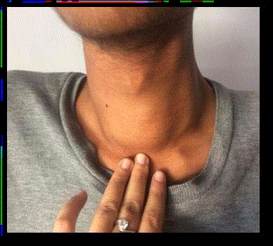
Figure 1
Pre-operative Clinical Photograph of Patient.
Figure 2
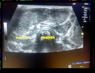
Figure 2
Grey scale USG image demonstates replacement of thyroid
parenchyma by hypoechoic collection showing moving internal echoes with
few intervening septae.
Figure 3
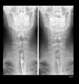
Figure 3
Barium swallow of the patient shows no abmormality. Bilateral
pyriform fossa are well delineated. No evidence of any pyriform sinus fistula
noted.
Figure 4
Figure 4
USG guided pus drainage.
Figure 5
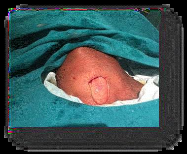
Figure 5
Intraoperative Incision &Drainage of Abcess.
Figure 6
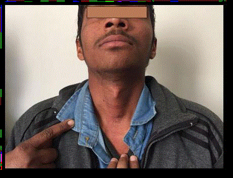
Figure 6
Follow up after 1 month.
Figure 7
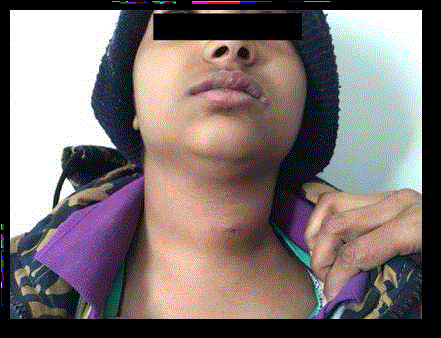
Figure 7
Pre-operative image showing swelling in the neck.
Figure 8
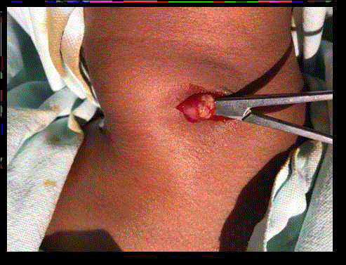
Figure 8
Intraoperative picture of the second patient with abscess.
Figure 9
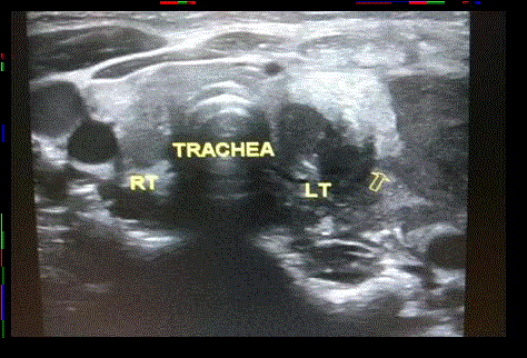
Figure 9
Grey scale USG image demonstates a heteroechoic area in left
lobe of thyroid communicating with a collection adjacent to left lobe of thyroid.
Surrounding fat planes were echogenic. Overlying strap muscles, skin and
subcutaneous tissue were slightly echogenic and edematous.
Figure 10
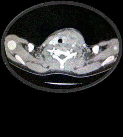
Figure 10
CECT neck of the patient shows a non-enhancing area in left
lobe of thyroid which is communicating with a heterogenously enhancing
collection in perithyroidal region. This is consistent with thyroid abscess with
contained rupture in perithyroidal region.
Discussion
Thyroid abscess and acute suppurative thyroiditis are not
common, representing only 0.1% to 0.7% of surgically treated thyroid
pathologies [1]. For this reason, diagnosis is often delayed, which
may lead to a life-threatening situation [2]. Rarity of this condition is
attributable to anatomic and physiologic characteristics of the glands
that impart a unique quality of infection resistance. These include
rich blood supply and lymphatic drainage, high glandular content of
iodine which can be bactericidal and separation of the gland due to
total encapsulation from other structures of neck [3].
Since the gland has no external connections the route of infection
was a mystery. Takai et al. [4] reported 15 patients with acute
supppurative thyroiditis where a piriform fistula was the apparent
route of infection. The pyriform sinus fistula is an internal pharyngeal
fistula and has been shown to be the most common underlying
abnormality in patients with AST. The fistula ends in or adjacent to
the thyroid and allows bacterial infection to develop in or around
the gland. The left side is more commonly involved than the right.
Thyroid abscesses usually start after upper respiratory tract, pharynx,
or middle-ear infections [5]. A case of thyrotoxicosis caused by acute
suppurative thyroiditis after repeated fine-needle aspiration (FNA)
has been described. Thyroid infection had possibly been induced by
needle-track seeding, because atopic skin favors colonization by S.
aureus because of local immunologic deficiency [6]. Rare cases such
as fish bone penetration through the esophageal mucosa into the
thyroid gland space of the neck after several weeks of swallowing has
been reported [7].
The most important causal organisms are Staphylococcus aureus,
Streptococcus species, and anaerobes. These infections account for
approximately 70% of cases [8]. Other causes include Escherichia coli
following urosepsis, Bacteroides fragilis in post-hysterectomy [9],
Klebsiella, Salmonella typhoid, Salmonella brandenburg, Eikenella
corrodens, Fusobacterium mortiferum [10], and aspergillosis [11].
Rare cases have been reported from Lemierre’s syndrome [12] (postanginal
septicaemia due to anaerobes) and infectious mononucleosis
in adolescence [13] presenting with thyroid abscess. Plain x ray may
show tracheal displacement and soft tissue swelling over thyroid
region. Barium swallow is mandatory to rule out pyriform sinus fistula
because it is a common cause of recurrent thyroid abscess and its total
resection effectively prevents a relapse. In the acute inflammatory
stage, USG showed a hypoechoic lesion spreading in and around the
affected thyroid lobe, destruction of the lobe, and abscess formation
in the neck. A careful review of the US studies demonstrated that the
following findings are characteristic of acute suppurative thyroidits:
a perithyroidal hypoechoic space, effacement of the plane between
the thyroid and perithyroid tissues, and the hypoechoic lesions being
unifocal. The former two are not seen in subacute thyroiditis, and
hypoechoic lesions in subacute thyroiditis are usually multiple and
often bilateral [9].
CT scans demonstrated similar features with clearer anatomical
involvement and edema in the ipsilateral hypopharynx. These findings
allowed easy diagnosis of AST. However, in the early inflammatory
stage USG showed an unclear hypoechoic area in the affected lobe
and CT scans showed a nonspecific low-density area. These findings
often led to erroneous diagnoses of subacute thyroiditis. In the late
inflammatory stage, USG and CT scans often showed atrophy and an
unclear hypoechoic or low-density area in and around the affected
lobe. To detect pyriform sinus fistulae, barium swallow studies are
more sensitive than USG or CT scans [9].
Role of FNAC is to differentiate acute suppurative thyroiditis and
subacute thyroiditis because the management lines are different for
these. It can also identify the bacteriological origin i.e. GPC or GNB
and thus helps to make a selective antibiotic selection [14].
References
- Menegaux F, Biro G, Schatz C, Chigot JP. Thyroid abscess: Apropos of 5cases. Ann Med Interne (Paris). 1991; 142: 99-102.
- Tien KJ, Chen TC, Hsieh MC, Hsu SC, Hsiao JY, Shin SJ, et al. Acute Suppurative Thyroiditis with Deep Neck Infection: A Case Report. Thyroid. 2007; 17: 467-469.
- Herndon MD, Christie DB, Ayoub MM, Duggan AD. Thyroid abscess: case report and review of the literature. Am Surg. 2007; 73: 725-728.
- Takai S, Miyauchi A, Matsuzuka F, Kuma K, Kosaki G. Internal fistula as a route of infection in acute suppurative thyroiditis. Lancet. 1979; 1: 751-752.
- Schweitzer VG, Nels R. Olson. Thyroid Abscess. Otolaryngol Head Neck Surg. 1981; 89: 226-229.
- Nishihara E, Miyauchi A, Matsuzuka F, Sasaki I, Ohye H, Kubota S, et al. Acute suppurative thyroiditis after fine-needle aspiration causing thyrotoxicosis. Thyroid. 2005; 15: 1183-1187.
- Ching Yuan Chen , Jyh Ping Peng. Esophageal fish bone migration induced thyroid abscess: case report and review of the literature. Am J Otolaryngol. 2011; 32: 253-255.
- Sicilia V, Mezitis S. A case of acute suppurative thyroiditis complicated by thyrotoxicosis. J Endocrinol Invest. 2006; 29: 997-1000.
- Yeluri S, Mehta JP, Karanth S, Guneesh Dadayal. A tender lump in the neck. MJA. 2006; 184: 137.
- Starvreas NP, Amanatidou, Emmanuel G. Hatzimanolis, Ioannis Legakis, George Naoum, Elli Lakka-Papadodima. Thyroid abscess due to a mixed anaerobic infection with fusobacterium mortiferum. J Clin Microbiol. 2005; 43: 6202-6204.
- Lisbona R, Lacourciere Y, Rosenthall L. Aspergillomatous abscesses of the brain and thyroid. J Nuc Med. 1973; 14: 541-542.
- Kara E, Sakarya A, Keles C, Borand H, Pekindil G, Göktan C. Case of lemierre’s syndrome presenting with thyroid abscess. Eur J Clin Microbiol. 2004; 23 : 570-572.
- Astl J, Kuchynkova Z, Taudy M. Thyroid abscess at an adolescent age. Int J Pediator Otorhinolaryngol. 2003; 67: 1375-1378.
- Paes JE, Burman KD, Cohen J, Franklyn J, McHenry CR, Shoham S. Acute bacterial suppurative thyroiditis: a clinical review and expert opinion. Thyroid. 2010; 20: 247-55.