Clinical Image
Indolent Intrahepatic Cholangiocarcinoma
Matteo Renzulli1*, Francesco Modestino1, Alberta Cappelli1, Cristina Mosconi1, Rita Golfieri1, Francesco Vasuri2, Antonietta D’Errico-Grigioni2, Giorgio Ercolani3 and Pietro Andreone4
1Department of Diagnostic Medicine and Prevention, S Orsola-Malpighi Hospital, University of Bologna, Italy
2Deparment of Oncology and Transplantation Pathology, S Orsola-Malpighi Hospital, University of Bologna, Italy
3Department of Medical and Surgical Sciences, S Orsola-Malpighi Hospital, University of Bologna, Italy
4Department of Medical and Surgical Sciences, Research Centre for the Study of Hepatitis, University of Bologna,
Italy
*Corresponding author: Matteo Renzulli, Department of Diagnostic Medicine and Prevention, Via Albertoni, 15, S Orsola-Malpighi Hospital, University of Bologna, 40138 Bologna, Italy
Published: 11 Jul, 2017
Cite this article as: Renzulli M, Modestino F, Cappelli
A, Mosconi C, Golfieri R, Vasuri
F, et al. Indolent Intrahepatic
Cholangiocarcinoma. Ann Clin Case
Rep. 2017; 2: 1397.
Clinical Image
A 58-year-old man, with hepatitis B virus-related cirrhosis, underwent magnetic resonance imaging (MRI) due to a >1 cm hypoechoic nodule identified during ultrasound surveillance. MRI demonstrated a 1.2 cm nodule suspected for intrahepatic cholangiocarcinoma (iCCA) (Figure 1). Laparoscopic hepatic resection was performed in a patient with good hepatic function (MELD 8, Platelet count 220^3/mmc, HVPG 6 mmHg and Fibroscan 6 kPa). At histology, the nodule was constituted of a ductal (pseudoglandular) proliferation, non-capsulated but with regular margins (image 10x, b) (Figure 2). Tumor cells showed mild cytological atypia and rare mitotic figures (image 20x, c), albeit the proliferative index (Ki67 immunohistochemistry, image 20x, c’) was increased (Figure 3). A diagnosis of well-differentiated iCCA (grade 1 according to WHO) was made. A retrospective reevaluation of an MRI performed more than 6 years before showed that the lesion was present and had a maximum diameter of 5 mm (d) (Figure 4). This is the proven evidence of a very early iCCA with an indolent natural history.
Figure 1
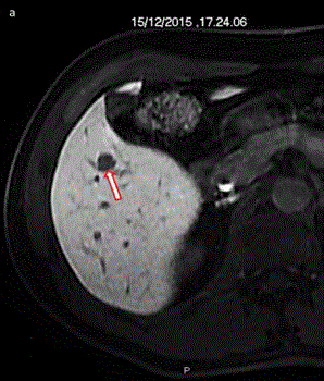
Figure 1
MRI demonstrated a 1.2 cm nodule suspected for intrahepatic cholangiocarcinoma.
Figure 2
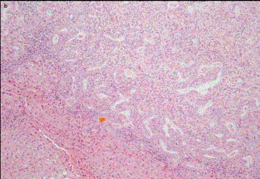
Figure 2
The nodule was constituted of a ductal (pseudoglandular) proliferation, non-capsulated but with
regular margins.
Figure 3
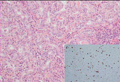
Figure 3
Tumor cells showed mild cytological atypia and rare mitotic figures.
Figure 4
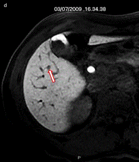
Figure 4
A retrospective reevaluation of an MRI performed more than 6
years before showed that the lesion was present and had a maximum
diameter of 5 mm.