Case Report
Vertebral Artery Dissection and Cerebellar Infarcts Presenting as a Typical Migraine
Korb A* and Sonnekus PH
Department of Internal Medicine, Private Practice, South Africa
*Corresponding author: Anneli Korb, Department of Internal Medicine, Private Practice, PO Box 44512 Linden 2104, South Africa
Published: 29 Jun, 2017
Cite this article as: Korb A, Sonnekus PH. Vertebral Artery
Dissection and Cerebellar Infarcts
Presenting as a Typical Migraine. Ann
Clin Case Rep. 2017; 2: 1387.
Abstract
We present a case of vertebral artery dissection presenting as an atypical migraine with key features including localised pain and transient ischaemic symptoms.
Keywords: Cervicocerebral arterial dissection; Vertebral artery
Introduction
Headache and neck pain are common presenting symptoms of Cervicocerebellar Artery Dissection (CAD). It is often accompanied by ischaemic symptoms and focal neurological signs. Arterial dissections account for approximately 2% of ischaemic strokes [1]. It is an important cause of stroke in younger patients. In an evaluation of 1008 consecutive ischaemic stroke victims aged 15 to 49 Putaala et al. [2] found CAD to be the underlying aetiolgy in 15% of patients, second only to cardioembolic causes.
Case Presentation
A 31 year old migraineur presented to the emergency department with a worsening left sided occipital headache, left sided posterior neck pain, vertigo and disequilibrium with falling to the left side. These symptoms evolved into paresthesia in the left face, arm, and leg as well as dysarthria which resolved spontaneously before his arrival to the emergency department. The symptoms were preceded by chronic occipital headaches and neck pain over a one month period leading up to his presentation to the emergency department. There was no history of overt neck trauma or chiropractic manipulation. However, he does play soccer on a regular basis. At the time of examination in the emergency department the patient only had left sided hemicrania. Physical and neurological examination was unremarkable and revealed no focal signs. Cerebellar tests were normal. An initial non-contrast CT Brain was ordered in view of new unexplained neurological symptoms and revealed multiple low density non-haemorrhagic lesions in the left cerebellar hemisphere and pons. No subarachnoid haemorrhage was demonstrated. Aspirin and enoxaparin were given as antithrombotic therapy to prevent embolism. T2 flare images and diffusion weighted MRI sequences showed acute non-haemorrhagic left sided cerebellar infarcts (Figure 1). This was followed by CT angiography which revealed a free floating thrombus in the horizontal part of the distal left vertebral artery. Contrast flowed around the thrombus with no current angiographic features of obstruction or dissection (Figure 2). In the absence of obvious angiographic evidence of arterial dissection we postulated that spontaneous CAD occurred and that the ensuing intramural thrombus opened distally resulting in thrombus formation in the vessel leading to the above ischaemic symptoms. In view of the presence of thrombus formation full anticoagulation was commenced with warfarin and aspirin was continued. Enoxaparin was discontinued once a therapeutic INR was reached. Hemicrania gradually subsided over 48 hours. The patient was discharged on Warfarin and aspirin and advised to avoid any contact sports chiropractic neck manipulation, and any activity that involves abrupt rotation and flexion-extension of the neck. The patient will be reassessed clinically and angiographically in 3 months. If symptom free and angiography reveals normal vasculature Warfarin will be discontinued and the maintenance therapy will include dual antiplatelet agents.
Figure 1
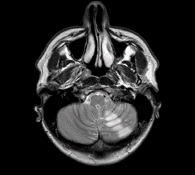
Figure 1
T2 flare images and diffusion weighted MRI sequences showed
acute non-haemorrhagic left sided cerebellar infarcts.
Figure 2
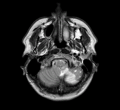
Figure 2
Contrast flowed around the thrombus with no current angiographic
features of obstruction or dissection.
Discussion
Spontaneous CAD is a relatively uncommon, but important cause of morbidity in young
patients presenting with suspected stroke or transient ischaemic events [1]. The reported annual
incidence has been estimated at up to 1.5 per 100,000 [3]. The etiology of spontaneous CAD is
largely unclear, but the following predisposing conditions have been repetitively implicated but
not systematically reviewed: migraine, hypertension, hereditary connective tissue disorders such as Ehlers-Danlos syndrome and Marfan syndrome, recent respiratory
tract infection, the use of oral contraceptive agents, hyperlipidaemia,
previous and current smoking, coagulopathies and substance abuse
[4-7]. Previous CAD is regarded an important risk factor with a
reported recurrence rate of 3-8% [8]. Possible pathogenetic factors
include hyperextension of the neck, particularly recent cervical
manipulation, as well as seasonal variation with a higher incidence
reported during autumn [9,10]. This variation is thought to be due to
an increase in respiratory tract infections during this period.
Common presenting symptoms include isolated headache,
posterior neck pain or both. A case series of 20 patients with sCAD
done by Arnold et al. [5] revealed that 30% presented with headache
only and 60% with headache and neck pain. The headache is often
described as a migraine with unusual features when compared to
previous migraines or not responsive to medication usually taken.
The headache is typically occipital, throbbing in nature and can be
ipsilateral to the dissection or bilateral. Other presenting symptoms
include dizziness, tinnitus, visual disturbances, balance disturbances
and limb weakness [7,9]. In a population-based outcome study Lee et
al. [11] demonstrated that 56% of patients with spontaneous cerebral
artery dissections presented with symptoms of cerebral ischaemia,
ranging from transient ischaemic attacks to signs of infarction. Other
signs of ischaemia may include retinal artery occlusion, ischaemic optic neuropathy and signs of posterior circulation infarction
syndromes and cervical myelopathy or radiculopathy [12,13]. A
proportion of patients can present with a subarachnoid haemorrhage
(SAH) as a result of intradural vertebral or basilar artery dissecting
aneurysm formation and subsequent rupture [14].
Especially in young patients presenting to the emergency
department with an atypical history or sings, the treating physician
must have a high index of suspicion for CAD. CT angiography
has a high sensitivity for the diagnosis of CAD and was previously
considered the “gold standard” for the diagnosis of CAD however MRI
and magnetic resonance angiography, that also poses a high sensitivity
by revealing the intramural haematoma and luminal stenosis or
occlusion, replaced the conventional CT angiogram. Transcranial
Doppler studies may reveal lesions in the vertebral arteries in the
neck. Thorough cardiovascular investigations, including a heart
sonar and electrocardiography are indicated to rule out cardiac and
aortic pathology. Evaluation for coagulopathies is also indicated. The
patient’s history will provide guidance for the indication of other
special investigations like screening for anti- phospholipid syndrome
and other genetic or acquired coagulopathies [7,15].
Several medical and surgical approaches are available for
the immediate management of CAD. A uniform approach is
lacking, most likely due to the absence of randomized trials. Most
importantly a subdural haemorrhage has to be excluded. Patients
presenting within 3 hours of initial presentation and not posing
a definite contra-indication for thrombolysis, the administration
of intravenous tissue plasminogen activator (t-PA) may enhance
neurological recovery. Optimal brain perfusion is important [7].
Endovascular vertebrobasiler angioplasty and stenting are technically
very difficult procedures and has a high complication rate but is used
if medical therapy is contra-indicated or has failed. Endarterectomy
for severe extracranial vertebral artery disease, when conducted by
an experienced surgeon, has a relative low complication rate [7,15,9].
Anticoagulation with intravenous heparin followed by Warfarin
anticoagulation with target International Normalized Ration (INR)
of 2-3 for three to six months is usually recommended although no
clear guidelines are available. Some authors recommend a follow up
MRI after three months and suggest a change to anti platelet therapy
if intraluminal irregularities are present [9].
Conclusion
Especially in young patients presenting to the emergency department with an atypical history or sings, the treating physician must have a high index of suspicion for CAD.
References
- Redekop GJ. Extracranial carotid and vertebral artery dissection: a review. Can J Neurol Sci. 2008; 35: 146-152.
- Putaala J, Metso AJ, Metso TM, Konkola N, Kraemer Y, Haapaniemi E, et al. Analysis of 1008 consecutive patients aged 15 to 49 with first-ever ischemic stroke: the Helsinki young stroke registry. Stroke. 2009; 40: 1195-1203.
- Bassetti C, Carruzzo A, Sturzenegger M, Tuncdogan E. Recurrence of Cervical Artery Dissection: A Prospective Study of 81 Patients. Stroke. 1996; 27: 1804-1807.
- Haneline MT, Rosner AL. The etiology of cervical artery dissection. J Chiropr Med. 2007; 6: 110-120.
- Arnold M, Cumurciuc R, Stapf C, Favrole P, Berthet K, Bousser MG. Pain as the only symptom of cervical artery dissection. J Neurol Neurosurg Psychiatry. 2006; 77: 1021-1024.
- Schievink WI, Michels VV, Piepgras DG. Neurovascular manifestations of heritable connective tissue disorders A review. Stroke. 1994; 25: 889-903.
- Savitz SI, Caplan LR. Vertebrobasilar Disease. N Engl J Med. 2005; 352: 2618-2626.
- Schievink WI, Mokri B, O'Fallon WM. Recurrent spontaneous cervical-artery dissection. N Engl J Med. 1994; 330: 393-397.
- Park KW, Park JS, Hwang SC, Im SB, Shin WH, Kim BT. Vertebral Artery Dissection: Natural History, Clinical Features and Therapeutic Considerations. J Korean Neurosurg Soc. 2008; 44: 109-115.
- Micheli S, Paciaroni M, Corea F, Agnelli G, Zampolini M, Caso V. Cervical Artery Dissection: Emerging Risk Factors. Open Neurol J. 2010; 4: 50-55.
- Lee VH, Brown RD Jr, Mandrekar JN, Mokri B. Incidence and outcome of cervical artery dissection: a population-based study. Neurology. 2006; 67: 1809-1812.
- Biousse V, Touboul PJ, D'Anglejan-Chatillon J, Levy C, Schaison M, Bousser MG. Ophthalmologic manifestations of internal carotid artery dissection. Am J Ophthalmol. 1998; 126: 565-577.
- Crum B, Mokri B, Fulgham J. Spinal manifestations of vertebral artery dissection. Neurology. 2000; 55: 304-306.
- Pozzati E, Andreoli A, Limoni P, Casmiro M. Dissecting aneurysms of the vertebrobasilar system: study of 16 cases. Surg Neurol. 1994; 41: 119-124.
- Thanvi B, Munshi SK, Dawson SL, Robinson TG. Carotid and vertebral artery dissection syndromes. Postgrad Med J. 2005; 81: 383-388.