Case Report
Is Preeclampsia Really an Uncommon Etiological Factor of Pleural Effusions?
Serdar Ozkan1* and Kerem Tetik2
1epartment of Thoracic Surgery, Medova Hospital, Turkey
2Department of Gynecology and Obstetrics, Cengiz Gökçek Gynecology and Obstetrics Hospital, Turkey
*Corresponding author: Serdar Ozkan, Department of Thoracic Surgery, Medova Hospital, Şeyh Şamil Mah. Dosteli cad.No:52/1, Selçuklu, Konya, Turkey
Published: 22 Jun, 2017
Cite this article as: Ozkan S, Tetik K. Is Preeclampsia
Really an Uncommon Etiological Factor
of Pleural Effusions?. Ann Clin Case
Rep. 2017; 2: 1384.
Abstract
Effusion developing due to preeclampsia in the pleural effusion etiology is quite rare, and in most studies, the term of preeclampsia is not even mentioned as to differential diagnosis. In the same way, pleural effusion is also rarely encountered in cases of preeclampsia in obstetry clinics. Transudative pleural pericardial effusion accompanied by ascites in an asymptomatic patient with mild preeclampsia that we diagnosed accidentally suggested that such cases could be more common than expected.
Keywords: Preeclampsia; Effusion; Ascites; Pleura
Introduction
Pleural effusion is a frequently seen entity in Turkey due to the diseases, such as congestive heart failure, malignancy, parapneumonic and pulmonary embolism, and tuberculosis. An increase in hydrostatic pressure, a reduction in oncotic pressure and an increased endothelial permeability are the most common mechanisms of pleural effusion [1]. Pleural effusion is encountered as a rare finding in cases with postnatal severe preeclampsia [2].
Case Presentation
A 31-year-old female patient with the history of previous and healthy pregnancy and delivery was admitted to the hospital for the second birth. Because her arterial pressure was measured as 210/100 mmHg on left and 160/100 mmHg on right arms on the first postnatal day, calcium channel blocker was started due to hypertension, and thorax and abdominal Computed Tomography (CT) were analyzed with the suspicion of aortic dissection. On CT investigation, pleuro-pericardial effusion and ascites were evaluated (Figure 1). No pathologic signs of aortic dissection were seen. In terms of the etiology of effusion, samples were obtained from pleural effusion through thoracentesis, and colorless and serous fluid was drained. In simultaneously performed pleural fluid/blood analyses, albumin, LDH and protein were measured as 0.22/2.22g/ dL, 74/1061 U/L and 0,47/5.01 mg/dL respectively, and transudate fluid was considered. No abnormality except for +2 proteinuria was encountered in the urine analysis. The patient in whom hematological and biochemical parameters were within normal limits except mild elevated transaminase was diagnosed with mild preeclampsia. Given that arterial pressure regressed to normal limits, the case was started to be treated with magnesium sulfate due to the prophylaxis of convulsion. On thoracic ultrasound performed on the second day, it was observed that no additional pathology was present, and pleuro-pericardial effusion was nearly ameliorated.
Figure 1
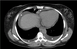
Figure 1
Chest CT imagerevealingpleural-pericardial effusion and ascites in
an asypmtomatic patient.
Discussion
The etiology of preeclampsia still remains unknown exactly, but several etiologies have
been proposed, including abnormal trophoblast invasion of uterine vessels, immunological
intolerance between fetoplacental and maternal tissues, maladaptation to cardiovascular changes
and inflammatory changes of pregnancy [3]. Preeclampsia involves wide spread endothelial cell
dysfunction. Massive fluid leakage into the third space occurs because of widespread endothelial
cell dysfunction [4].
As a result of the investigations performed in our case, the fluid detected in abdomen and
pleuro-pericardial space was considered to be transudative. When analyzing the factors leading to
transudative fluid, the decrease seen at serum protein and albumin levels in our case was thought to
be the primary cause of effusion. However, endothelial dysfunction responsible preeclampsia is also considered to be involved in the etiology of effusion.
In our case, the presentation of mild preeclampsia was assumed
in light of clinical and laboratory signs, except for arterial pressure
of 100 mmHg. According to Cong [5], although the incidence of
ascites was found to be 21.6/1000 in severe cases of preeclampsia, the
development of pleuro-pericardial effusion as mild preeclampsia is
the sign of a rare condition.
The diagnosis of effusion was performed incidentally in our case
due to lack of symptoms such as angina, cough or shortness of breath
dispnea. The fact that the effusion leads to no pleural-peritoneal
irritation and formation of other symptoms because of its poor
content may be encountered as normal, but non-formation of dispnea
due to pleural effusion and non-occurrence of cardiac symptoms due
to pericardial effusion should be assessed as conditions requiring
further studies.
We wish to emphasize once more that preeclampsia should be
kept in mind in the differential diagnosis of the effusion followed-up
in women with pregnancy. We consider that the rarity of effusion
incidence due to preeclampsia in thoracic surgery and obstetrics may
be a relative decrease because of the asymptomatic courses of such
cases.
References
- Rashid RJ, Jalili J, Arhami SS, Heris HK, Habibzadeh A, Mohtasham MA. The accuracy of chest computed tomography findings in differentiation of exudative from transudative pleural effusion. J Clin Anal Med. 2013.
- Kumar R, Dey M. Massive ascites and bilateral pleural effusion causing respiratory embarrassment in a postnatal case of severe preeclampsia. Med J Armed Forces India. 2014; 70: 290-292.
- Sibai B, Dekker G, Kupferminc M. Pre eclampsia. Lancet. 2005; 365: 785-799.
- Brown MA, Zammit VC, Lowe SA. Capillary permeability and extracellular fluid volumes in pregnancy-induced hypertension. Clin Sci. 1989; 77: 599-604.
- Cong KJ, Wang TT. Complication of ascites in pregnancy induced hypertension. Zhonghua Fu Chan Ke Za Zhi. 1994; 29 :7-9.