Case Report
A Rare Case of Laryngeal Schwannoma
Bhavna B Kamble*, Prasad Deshmukh, Disha Methwani and Puja Lakhotia
Department of ENT, Jawarharlal Nehru Medical College Sawangi (M), India
*Corresponding author: Bhavna B Kamble, Department of ENT, Jawarharlal Nehru Medical College and A.V.B.R.Hospital Sawangi (M), Wardha, (442001) Maharashtra, India
Published: 05 Jun, 2017
Cite this article as: Kamble BB, Deshmukh P, Methwani D,
Lakhotia P. A Rare Case of Laryngeal
Schwannoma. Ann Clin Case Rep.
2017; 2: 1364.
Abstract
Introduction: Schwannoma within larynx is uncommon and arises from perineural schwann cells. These tumors are a potential threat to airway and usually cause a challenge to otorhinolaryngologists in diagnosis and management. A definite diagnosis can only be made histologically. Very few
benign lesions due to location, size and site of origin need total or partial laryngectomy. Laryngeal
Schwannoma is one of them. We present a case report of this rare laryngeal entity wherein diagnosis
was tortuous and tormenting and various investigations gave conflicting information adding to the
diagnostic dilemma.
Methods: A case report of laryngeal schwanomma in a young patient in a tertiary care rural hospital
in September 2015 is discussed
Result/Discussion: The clinical course, quest of diagnosis, conflicting inputs given by various
investigations, management is discussed.
Conclusions: Our case is unique as laryngeal schwanomma is a rare entity with diagnostic crisis.
Decision making for management was also challenging due to benign nature and extent of lesion.
In light of final histopathology report agreeing with USG guided report, we feel later should be
considered as a reliable implement for diagnosis of this rare tumour.
Keywords: Laryngeal schwannoma; Stridor; Neurilemoma
Introduction
Schwannoma within larynx is uncommon and consists of only 0.1% to 1.5% of all benign
laryngeal tumours [1,2]. As few as 250 cases are reported in literature till date [3]. Schwannoma arises from perineural schwann cells and are well encapsulated, slow growing, submucosal abutting the parent nerve but extrinsic to nerve fascicles [1,4]. Around 80% are located in the region of aryepiglottic folds and 20% are located in region of false and true vocal cords [4] and known to arise usually from internal branch of superior laryngeal nerve [2,4].
Laryngeal schwannomas usually occur in females in 4th and 5th decades. These tumors are a potential threat to airway and usually cause a challenge to otorhinolaryngologists in diagnosis and management [5]. A definite diagnosis can only be made histologically [6]. The most successful curative option is complete surgical resection [7]. Scores of articles with detailed literature review of Laryngeal Schwannoma have been published. Practising Otorhinolaryngologists should always be alive to the possibility of this uncommon laryngeal lesion. Very few benign lesions due to location, size and site of origin need total or partial laryngectomy. Laryngeal Schwannoma is one of them. We
present a case report of this rare laryngeal entity.
Case Presentation
A 27 years old female came to ENT OPD with history of change in voice since a year. Onset
of hoarseness of voice was sudden without any precipitating factors like upper respiratory tract
infection, trauma, surgical procedure and it worsened gradually for the past one year. Symptom
was refractory to all the medications that she took from general practitioners. Around same time
she also developed mucoid expectoration and breathlessness on exertion. Peculiar symptom of dull
pain on the left side of neck was characteristic .There was no fever, dysphagia, odynophagia, neck
swelling or stridor. Patient had no addiction and no known systemic illness.
On Indirect laryngoscopy, there was submucosal swelling involving left areyepiglottic fold,
areytenoids, left vocal cords and extending to left pyriform fossa. Patient was admitted, investigated
and a direct laryngoscopy under general anaesthesia with biopsy was planned. Direct laryngoscopy
using Chevalier Jackson type of direct laryngoscope was performed. During direct laryngoscopy,
a smooth, firm, submucosal mass arising from the left hemilarynx was seen and biopsy was performed. At the end of the procedure patient suddenly developed
stridor. for which emergency tracheostomy was done and a cuffed 7.5
portex tracheostomy tube inserted. Air blast was good but following
tracheostomy patient had developed emphysema over face, neck,
chest. An emergency chest radiogram posterior anterior view revealed
bilateral pneumothorax (Figure 1) which was taken care by putting
Intercostal Drain no 28 in 4th intercostal space in midclavicular line
for 3 days (Figure 2). Patient responded positively and was stable.
The biopsy report was inconclusive showing only squamous
cell hyperplasia. Patient was further investigated. Ultrasonography
was suggestive of possible Schwanomma or leiomyoma whereas
Computed Tomogram scan uncovered heterogeneously enhancing
soft tissue density mass lesion in hypopharynx on left side involving
left side of cricoid extending upto the right half of cricoid cartilage
with near obliteration of tracheal lumen (Figure 4 and 5) most likely
to be neoplastic.
Patient was re-biopsied but report was far from conclusive and
showed chronic lymphoplasmacytic inflammatory infiltrate with no malignant cells. With no diagnosis forthcoming from above
investigations Ultrasound guided Fine Needle Aspiration Cytology
was performed which was reported as Schwannoma and a deeper
tissue biopsy was advised for confirmation (Figure 3). Accordingly
patient was subjected to third biopsy and this time reported as
Enchondroma.
Though the lesion was benign its location, site, extent and
possible origin from cricoids with histopathology report as
enchondroma prompted us perform a total laryngectomy. Tracheal
stoma was created by taking bevelled shaped incision at 2nd tracheal
ring. Neopharynx was created in a T shaped manner after preserving
maximum right pyriform fossa mucosa. A 7.5 no portex cuffed
tracheostomy tube and Ryle’s tube number 16 was inserted at the
time of creation of neopharynx.
The mass was firm globular to ovoid of about 5 cm x 5 cm x 4 cm
in dimensions arising from the left Areepiglottic folds extending to
and involving the left vocal cord, anterior commisiure and cricoids
causing bulge into pyriform fossa on the left side pushing epiglottis
to the right side. No involvement of trachea was observed (Figure 6).
Patient was kept on broad spectrum antibiotics. Nebulisation
with budacort, salbutamol and mucomix was given. Meticulous
stomal care and wound dressing was done. Drain was removed on 3rd
postoperative day. Ryle’s feeding was started on 3rd postoperative day
and continued upto 21 days. On 14th day oral feeds were started after
ruling out pharyngocutaneous fistula. There were no perioperative
complications. All in all patients made a remarkable recovery and
discharged on 23rd postoperative day with a permanent healthy stoma.
Patient was followed up after 15 days. Tissue was fixed in formalin for 48 hrs and sectioned in 3 mm slices. Final histopathology report
was schwanomma of left recurrent laryngeal nerve showing spindle
nuclear cells placed in palisades appearing like a verocay body (Figure 7) Patient is under follow up and doing well. We are planning for a
secondary q234 Tracheosophageal Prosthesis for voice rehabilitation
in coming few months.
Figure 1
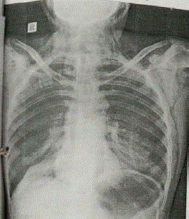
Figure 1
Chest radiogram postero-anterior view showing pneumothorax.
Figure 2
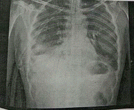
Figure 2
Radiogram after insertion of Intercostal Drain.
Figure 3
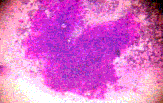
Figure 3
Ultrasound guided Fine Needle Aspiration Cytology suggestive
possibility of schwannoma or leiomyoma.
Figure 4
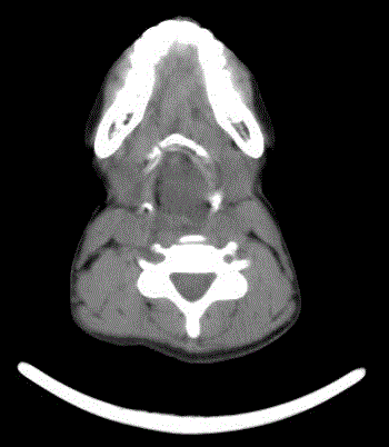
Figure 4
Computed tomogram showing the extent of tumour.
Figure 5
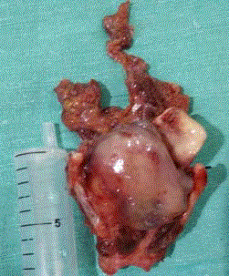
Figure 5
Total laryngectomy specimen showing tumour.
Figure 6
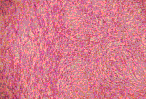
Figure 6
Postoperative histopathology showing schwanomma.
Figure 7

Figure 7
Stoma at the time of discharge.
Discussion
Schawannoma is a slow growing, benign, encapsulated tumour
arising from schwann cells. Laryngeal schwannoma is extremely rare
and most commonly found in females in 4th to 5th decade of life [8].
However no age is immune to this entity. The case we are reporting
is also a young female in her 3rd decade. Most commonly involved is
the internal branch of superior laryngeal nerve [9,10]. Often however,
this is not noticeable intraoperatively and likely to originate from the
smaller distal nerve fibres in the laryngeal submucosa [10]. In this
case too, parent nerve was not discernible. Schwannomas classically
affect nerve sheaths rather than nerve fibres and hence symptoms are
determined by site and mass effect [11,12].
Laryngeal schwannomas may approach a large size, causing
upper airway obstruction, dysphonia and even vocal cord fixation,
depending on their location, however most of them have insidious
course [13-16]. Two distinct feature in our case were sudden
hoarseness of voice and dull pain. It is said that dull pain is very
typical of schwannoma [9].
Most common site is at areyepiglottic folds (80%), areytenoids,
ventricular folds and true and false vocal cords (20%) [17,18]. The
diagnostic work-up includes indirect laryngoscopy which usually
reveals a submucosal mass. CT findings for benign laryngeal
schwannoma usually include heterogenous enhancement of the
lesion, no cartilage erosion and absence of infiltrative pattern [16].
The lesion is usually round to oval is attenuated with muscle and
sharply demarcated [18]. In this case site of origin was AE folds. In the quest of diagnosis, CONFLICTING inputs GIVEN BY VARIOUS
investigations led to diagnostic impasse. Initially two biopsy were
inconclusive while third biopsy was reported as enchondroma.
Neoplastic etiology was suspected on Computes Tomography scan.
Ultrasound and Ultrasound guided Fine Needle Aspiration Cytology
revealed a diagnosis of Schwannoma. Variable opinions of various
investigations compounded the diagnostic process. There are many
cases in literature suggesting that Fine Needle Aspiration Cytology
has low accuracy in the diagnosis of neural tumours [19-21]. But it was
Ultrasound & Ultrasound guided Fine Needle Aspiration Cytology
report which concured with final histopathology report in our case.
The preoperative diagnosis may be difficult. Intraoperative findings
help us to finalize our diagnosis [12]. Enger and Weiss established
three histological criteria for the diagnosis of schwannoma:
encapsulation, presence of Antoni A and/or Antoni B stroma, and
S-100 protein positivity [22].
Malignant transformation is rare in Schwanomma and is highly
radioresistant [23]. Complete surgical resection is the treatment of
choice. Depending upon the site size and location, a median or lateral
thyrotomy or a median or lateral pharyngotomy are the various
surgical approaches especially for tumours more than 5 cm. For
smaller tumor, endoscopic endolaryngeal CO2 laser assisted excision
can also be done [1,4,24,25]. Above procedures should be done
without disturbing the laryngeal framework.
In our case the mass was involving left areyepiglottic fold,
left areytenoid extending to left side cricoid cartilage and also
crossing midline anteriorly. As any partial procedure would have
made the laryngeal framework highly unstable, decision in favour
of total laryngectomy was made. Our case is unique as laryngeal
schwanomma is a rare entity, diagnosis was tortuous and tormenting
and various investigations gave conflicting information adding to
the diagnostic dilemma. Decision making for management was also
challenging due to benign nature and extent of lesion. In light of final
histopathology report agreeing with USG guided report, we feel later
should be considered as a reliable implement while sailing through
such a diagnostic crisis.
References
- Chiu CC, Chou SH, Wu CC, Liang PI, Lee KW. Obstructive laryngeal schwannoma in a young female. World J Surg Oncol. 2015; 13: 24.
- López-Álvarez F, Gómez-Martínez JR, Suárez-Nieto C, Llorente-Pendás JL. Schwannoma of the larynx. An infrequent laryngeal tumour. Acta Otorrinolaringol Esp. 2013; 64: 157-160.
- Boros MJ, Wysong ST. Syndromes after resection of cervical schwannoma. Ear Nose Throat J. 2011; 90: 431-433.
- Rao SM, Chandra ST, Kumar AY, Murthy PSN. Laryngeal schwannoma excision by lateral pharyngotomy: a case report. IJOPL. 2011; 1: 37-39.
- Vital I, Fliss DM, Cohen JT. Laryngeal schwannoma excised under direct laryngoscopy: Case report. Ear Nose Throat J. 2012; 91: 204-205.
- Ebmeyer J, Reineke U, Geh HB, Hamberger U, Mlynski R, Essing M, et al. Schwannoma of the larynx. Head Neck Oncol. 2009; 1: 24.
- Wang B, Dong P, Shen B, Xu H, zheng J. Laryngeal schwannoma excised under a microlaryngoscope without tracheotomy: A case report. Exp Ther Med. 2014; 7: 1020-1022.
- Ramakrishnan Y, Issing WJ. Laryngeal Schwannoma: Case Report and Literature Review. ISRN Otolaryngology. 2011; 2011: 1-3.
- Arora N, Jain K, Bansal R, Passey JC. Laryngeal schwannoma-A rarely occurring benign tumor. Otolaryngology Online J. 2015; 5: 5.
- Nanson EM. Neurilemoma of the larynx: a case study. Head and Neck Surgery. 1978; 1: 69–74.
- Tse A, Anwar B. Laryngeal schwannoma: excision via a laryngofissure approach. J Surg Case Rep. 2015; 2015: rjv059.
- Tas E, Vural S, Cuhali B, Turkoz H, Gursel A. Extracranial Head And Neck Schwannomas. The Internet Journal of Head and Surgery. 2013; 2.
- Saita V, Azzolina A, Galia A, Fraggett F. Schwannoma of epiglottis: A case mainly focusing on clinicopathological aspects. Acta Otorhinolaryngol Ital. 2005; 25: 378–380.
- Cadoni G, Bucci G, Corina L, Scarano E, Almadori G. Schwannoma of the larynx presenting with difficult swallowing. Otolaryngol Head Neck Surg. 2000; 122: 773-734.
- Bozec A, Dassonville O, Poissonnet G, Ndiaje M, Demard F. Laryngeal schwannoma: a case report. Ann Otolaryngol Chir Cervicofac. 2003; 120: 40-44.
- Tzagkaroulakis A, Stivaktakis J, Nikolopoulos T, Davilis D, Zervoudakis D. Ancient schwannoma of the true vocal cord. ORL J Otorhinolaryngol Relat Spec. 2003; 65: 310-313.
- Palva T, Jokinen K, Karja J. Neurilemmoma (schwannoma) of the larynx. J Laryngol Otology. 1975; 89: 203–207.
- Rosen FS, Pou AM, Quinn FB. Obstructive supraglottic schwannoma: a case report and review of the literature. Laryngoscope. 2002; 112: 997-1002.
- Bocciolini C, Dall'olio D, Cavazza S, Laudadio P. Schwannoma of cervical sympathetic chain: Assessment and management. Acta Otorhinolaryngol Ital. 2005; 25: 191-194.
- Hood RJ, Reibel JF, Jensen ME, Levine PA. Schwannoma of the cervical sympathetic chain. The Virginia experience. Ann Otol Rhinol Laryngol. 2000; 109: 48-51.
- Colreavy MP, Lacy PD, Hughes J, Bouchier-Hayes D, Brennan P, O'Dwyer AJ, et al. Head and neck schwannomas - a 10 year review. J Laryngol Otol. 2000; 114: 119-124.
- Kleihues P, Cavenee WK, Woodruff JM, Kourea HP, Louis DN, Schethauer DW. Schwannoma in WHO Classification of Tumours: Pathology and Genetics of Tumorus of Nervous System Eds., pp. 164–166, IARC Press, Lyon, France, 2nd edition, 2000.
- Sofi FA, Mir MH, Bagdadi FS, Mehmood K. Hidden Diagnosis in the Subglottic Larynx: Schwannoma Mimicking as Bronchial Asthma. N Am J Med Sci. 2012; 4: 325–327.
- Kun Z, Qi DY, Zhang KH. A comparison between the clinical behaviour of neurilemmomas in the neck and oral and maxillofacial region. J Oral Maxillofac Surg. 1993; 51: 769-719.
- Lin J, Martel W. Cross-sectional imaging of peripheral nerve sheath tumors: characteristic signs on CT, MR imaging, and sonography. AJR Am J Roentgenol. 2001; 176: 75-82.