Case Series
Interventional Management of Chronic Low Back Pain Associated with Bertolotti's Syndrome: Report of Case Series
Rui Zhang* and Jianguo Cheng
Department of Pain Management, Anesthesiology Institute, Cleveland Clinic, Ohio, USA
*Corresponding author: Rui Zhang, Departments of Pain Management, Anesthesiology Institute, Cleveland Clinic, 9500 Euclid Avenue/ C25, Cleveland, Ohio 44195, USA
Published: 04 May, 2017
Cite this article as: Zhang R, Cheng J. Interventional
Management of Chronic Low Back Pain
Associated with Bertolotti's Syndrome:
Report of Case Series. Ann Clin Case
Rep. 2017; 2: 1348.
Abstract
Background: Bertolotti's syndrome is a congenital variant with an enlarged transverse process of
the L5 lumbar vertebra (lumbosacral transitional vertebra, LSTV), which articulates or fuses with
the sacrum or ilium. It’s believed that the presence of an LSTV increases the incidence of chronic
low back pain and the severity of the pain. Its association with chronic low back pain is multifactorial,
including biomechanical changes resulting from asymmetrical motion between the LSTV
and the sacrum. The conventional conservative management that consists of physical therapy,
activity modification, and medication may not be sufficient to provide satisfactory pain control due
to the underlying congenital anatomical variance. Here we report 5 cases that were managed with
interventional procedures for chronic low back pain associated with Bertolotti’s syndrome.
Case Report: All the cases have failed to respond to conventional conservative management and
were found to have Bertolotti’s syndrome through radiographs. The pain generating mechanism
varied among these five patients, which led to diverse clinic presentations and applications of various
interventional procedures including epidural steroid injections, lumbar facet medical branch block
and neurotomy, and sacroiliac joint injection and neurotomy.
Conclusion: In light of the diversity of clinical presentations, management of patients with
Bertolotti’s syndrome has to be highly individualized. Multimodal care, including interventional
approaches, is often required based on clinical data, imaging, and diagnostic blocks.
Introduction
Bertolotti's syndrome refers to a congenital variant with an enlarged transverse process of
the L5 lumbar vertebra (lumbosacral transitional vertebra, LSTV), which articulates or fuses with
the sacrum or ilium. Its correlation with low back pain was first described in 1917 by the Italian
physician Mario Bertolotti. The prevalence of Bertolotti's syndrome was estimated ranging from 4.6
-15.8% in the general population but increases up to 35.6% in those seeking care for low back pain
[1-5]. It’s also believed that LSTV increases the severity of patient’s clinical presentation and pain
[6]. Unilateral LSTV is more common than bilateral LSTV (9% vs. 3%) [4]. Men are more likely to
be affected than women (28.1% vs. 11.1%) [7]. Bertolotti syndrome is usually diagnosed based on
imaging studies, such as lumbosacral plain x-rays, Computed Tomographic (CT) scans, and Magnetic
Resonance Imaging (MRI). Its presence on the imaging study does not predict the symptoms of low
back pain. However, the presence of an LSTV often leads to increased incidence of low back pain
due to reduced and asymmetrical motions between the LSTV and the sacrum. The asymmetrical
motion can result in early arthritic changes or nerve root compression at the pseudoarticulation.
The restricted motion between the LSTV and the sacrum can lead to compensatory hypermobility
of the adjacent segments, causing facet pain or irritation of the nerve roots at that level, or sacroiliac
dysfunction [8,9]. Due to its biomechanical and pathophysiological changes, the pain generators can
be distinct in the patients who presented with low back pain associated with Bertolotti’s syndrome,
which imposes a challenge for successful management of these patients.
The conventional conservative management that consists of physical therapy, activity
modification, medication such as NSAIDs, muscle relaxants, anticonvulsants, and antidepressants
may not be sufficient to provide satisfactory pain relief. There is a dearth of literature regarding
interventional approaches to the management of patients with low back pain associated with
Bertolotti’s syndrome [8,10]. In this report, we describe five patients with Bertolotti’s syndrome who presented to a tertiary pain center with low back pain and/or radicular
leg pain after having failed conventional conservative management.
The pain generating mechanisms varied among these five patients,
which led to diverse clinic presentations. Different interventional
procedures were applied to achieve therapeutic effects.
Case Presentation
Case 1
A 52 year-old man presents with chronic low back pain for over 3
years. The pain was described as deep, dull and radiating to the gluteal
areas. The pain was exacerbated by walking and mitigated by heat,
physical therapy, massage and a TENS unit. There were no significant
neurological findings on physical exam. Radiographs demonstrate
bilateral pseudoarticulation and/or fusion between the L5 transverse
process and the ilium (Figure 1). A bilateral L3-L5 lumbar facet
medial branch radiofrequency neurotomy did not provide adequate
pain relief or functional improvement. But a L4-L5 interlaminar
epidural steroid injection using a mixture of lidocaine 1% and
corticosteroids provided 50% pain reduction for about 2.5 months.
The pain likely originated from nerve root irritation by the enlarged
transverse processes or pseudoarticulation. This patient currently gets
L4-L5 interlaminar epidural steroid injections every 3 months with
adequate pain control and functional improvement.
Case 2
A 57 year-old man presents with chronic right leg pain radiating
to the right dorsal foot for four years. The pain was described as a
constant burning sensation associated with numbness and tingling.
The pain was exacerbated by standing and getting up from a sitting
position and alleviated by lying down. The straight leg raise test was
positive on the right side and the tactile sensation was decreased on
the L5 distribution on physical exam. The lumbar spine MRI shows
evidence of a protruding disc at L4-L5, compressing and effacing
the L5 nerve root on the right side. Radiographs demonstrate fusion
between the left L5 transverse process with the ilium. Right L4 and L5
diagnostic and therapeutic transforaminal epidural steroid injection
using a mixture of lidocaine 1% and corticosteroids provided 60%
of immediate pain reduction after the procedure (Figure 2). Further
follow-up of outcomes is pending. His right lower leg and foot pain
could result from the lumbar radiculopathy caused by L4-L5 disc
herniation, which is common in patients with Bertolotti’s syndrome
in compensation to the limited range of motion around the L5-S1 intervertebral disc.
Case 3
A 65 year-old man presents with predominantly axial low back
pain and pain in the left posterior thigh for over 5 years. The pain
was described as aching and worse when getting up out of a sitting
position. Physical exam showed tenderness on deep palpation over the
bilateral facet joints, left worse than right, with the negative straight
leg raising test. Radiographs showed fusion of the left transverse
process of LSTV with the sacrum or ilium (Figure 3). Lumbar spine
MRI showed multilevel degenerative changes, transitional S1 with
moderate left lateral recess narrowing at L5-S1 and likely chronic
impingement of the left S1 nerve root. Left lumbar facet medial
branch radiofrequency neurotomy at 80 degrees for 90 seconds after
two previous positive diagnostic blocks provided greater than 60%
pain reduction. In this case, the lumbar facet arthropathy in the
segments above the sacrum likely contributed to his back pain. This is
probably from the hypermobility of these segments secondary to the
restricted motion between the LSTV and the sacrum. Unfortunately,
this patient was injured in a motor vehicle accident a few weeks after
the procedure and we have not been able to further follow up his
outcomes of the procedure.
Case 4
A 47 year old man presents with left low back pain radiating to
the posterior aspect of the left lower extremity down to the level of the
knee for 6 years. The pain was described as sharp and stabbing without numbness or tingling. Physical exam was positive for the straight leg
raise on the left side. He completed a course of physical therapy and
underwent bilateral lumbar medial branch blocks without significant
benefit. Lumbar spine radiographies showed asymmetric enlargement
of L5 transverse processes fused or articulate with the sacrum. The
pseudoarticulation between the left transverse process and the ilium
appeared more mobile than the one on the right side (Figure 4A).
He had 75-90% of pain reduction for duration of 2-7 months to each
left L5 transforaminal epidural steroid injection using a mixture of
lidocaine and corticosteroid (Figure 4B).
Case 5
A 62 year old man presents with right low back pain radiating
to the right gluteal area for 5years. The pain was described as aching
and worse with walking. The Patrick test was positive on the right
side. Lumbar spine radiographs demonstrated asymmetrical
pseudoarticulation and/or fusion between the L5 transverse processes
and the iliums (Figure 5A). This patient had excellent responses to
right sacroiliac joint injections with a mixture of corticosteroid and
local anesthetics (Figure 5B). Subsequently, denervation of the right
sacroiliac joint sensation via L5 dorsal ramus, S1 and S2 lateral branch
radiofrequency neurotomy provided 70-80% of pain reduction for
four months and he is still benefiting from the procedure (Figure
5C).The restricted motion between the LSTV and the sacrum may
have led to relative hypermobility and dysfunction of the adjacent
segments, including the sacroiliac joint in his case.
Figure 1
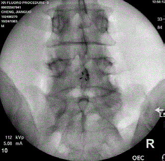
Figure 1
Anteroposterior radiographof case 1 demonstrates bilateral
pseudoarticulation and/or fusion between the enlarged L5 transverse
processes and the ilium and sacrum.
Figure 2
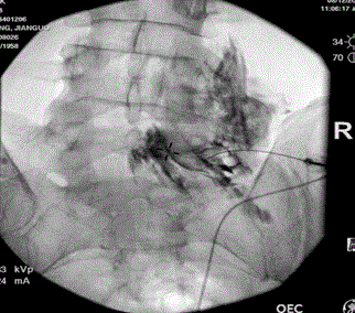
Figure 2
Anteroposterior radiographof case 2 demonstrates fusion between
the enlarged left L5 transverse process with the ilium and sacrum. L5
transforaminal epidural steroid injection was performed after confirmation
ofconstrast spreads in the epidural space and along the right L4 and L5
nerve roots.
Discussion
We describe five patients with Bertolotti’s syndrome who presented to a tertiary pain center with low back pain and/or radicular pain after having failed conservative treatments. Their clinical presentations and primary pain generators were different as discussed above in each individual case presentation. The interventional procedures that provided greater than 50% pain reduction in our study include L4- L5 interlaminar epidural steroid injection, L4 and L5 transforaminal epidural steroid injection, lumbar facet medial branch blocks and radiofrequency neurotomy, sacroiliac joint injections and subsequent L5 dorsal ramus, S1 and S2 lateral branch radiofrequency neurotomy. Management of chronic low back pain associated with Bertolotti’s syndrome should be initiated with a comprehensive conservative approach consisting of activity modification, physical therapy and home exercise, psychosocial support, and medications such as NSAIDs, muscle relaxants, anticonvulsants, and antidepressants. Interventional approaches can be beneficial in cases refractory to conventional conservative management. For those who presented with lumbosacral radiculopathy or radiculitis symptoms due to the direct compression or irritation of the nerve root by an enlarged transverse process or pseudoarticulation, a transforaminal or interlaminar epidural steroid injection maybe considered. A diagnostic lumbar facet medial branch block can be performed for those with clinical evidence of lumbar facet pain due to the reduced motion between the LSTV and the sacrum. Radiofrequency neurotomy of the target medial branches may provide longer-term pain relief if the patient has greater than 50% pain reduction from the diagnostic medical branch block. For those suspected of sacroiliac dysfunction due to increased load to the sacroiliac joint with restricted motion between the LSTV and the sacrum, a diagnostic sacroiliac joint injection maybe attempted. If the patient has greater than 50% pain reduction, radiofrequency neurotomy of the L5 dorsal ramus, S1 and S2 lateral branches may provide longer-term pain relief. A few other interventional procedures to manage chronic low back pain associated with Bertolotti’s syndrome have also been reported. Injections of steroids and/or local anesthetics into the LSTV pseudoarticulation site can be both diagnostic and therapeutic in providing temporary pain relief if the primary pain generator is the pseudarthrosis [11]. Posterolateral fusion, partial or full resection of the LSTV pseudoarticulation have been reported to provide pain reduction and can be considered for long-term relief if the diagnostic block of pseudoarticulation was positive [12-14].
Figure 3
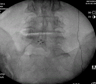
Figure 3
(a) Lumbar spine radiographof case 4 (from a previus left
medial branch block procedure) shows asymmetric enlargement of L5
transverse processes fused or articulate with the sacrum and/or ilium. The
pseudoarticulation between the left transverse process and the ilium appears
more mobile than the one on the right side. (b) L5 transforaminal epidural
steriod injection was performed after confirmation of constrast spreads in the
epidural space and along the right L5 nerve root.
Figure 4
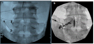
Figure 4
(a) Lumbar spine radiographof case 4 (from a previus left
medial branch block procedure) shows asymmetric enlargement of L5
transverse processes fused or articulate with the sacrum and/or ilium. The
pseudoarticulation between the left transverse process and the ilium appears
more mobile than the one on the right side. (b) L5 transforaminal epidural
steriod injection was performed after confirmation of constrast spreads in the
epidural space and along the right L5 nerve root.
Figure 5
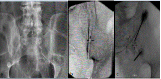
Figure 5
a) Lumbar spine radiograph of case 5 demonstrates asymmetrical
pseudoarticulation and/or fusion between the L5 transverse processes and
the iliums. b) Right sacroiliac joint injection. c) L5 dorsal ramus, S1 and S2
lateral branch radiofrequency neurotomy.
Conclusion
In light of the diversity of clinical presentations, management of patients with low back pain associated with Bertolotti’s syndrome has to be individualized. Multimodal care is often required based on clinical data, imaging and diagnostic blocks. Interventional procedures may provide better pain relief and facilitate participation in functional physical activities. Understanding the biomechanical and pathophysiological mechanisms underlying the chronic low back pain is the key to choosing the appropriate procedures.
References
- Apazidis A, Ricart PA, Diefenbach CM, Spivak JM. The prevalence of transitional vertebrae in the lumbar spine. Spine J. 2011;11(9):858-62.
- Quinlan JF1, Duke D, Eustace S. Bertolotti's syndrome. A cause of back pain in young people. J Bone Joint Surg Br. 2006;88(9):1183-6.
- Tang M, Yang XF, Yang SW, Han P, Ma YM, Yu H, et al. Lumbosacral transitional vertebra in a population-based study of 5860 individuals: prevalence and relationship to low back pain. Eur J Radiol. 2014;83(9):1679–82.
- Mahato NK. Morphometric analysis and identification of characteristic features in sacra bearing accessory articulations with L5 vertebrae. Spine J. 2010;10(7):616–21.
- Delport EG, Cucuzzella TR, Kim N, Marley J, Pruitt C, Delport AG. Lumbosacral transitional vertebrae: incidence in a consecutive patient series. Pain physician. 2006;9(1):53–6.
- Taskaynatan MA, Izci Y, Ozgul A, Hazneci B, Dursun H, Kalyon TA. Clinical significance of congenital lumbosacral malformations in young male population with prolonged low back pain. Spine. 2005;30(8):E210-E13.
- Nardo L, Alizai H, Virayavanich W, Liu F, Hernandez A, Lynch JA, et al. Lumbosacral transitional vertebrae: association with low back pain. Radiology. 2012;265(2):497–503.
- Wu J, Cheng J. Unusual pain syndromes. Kaye A, Shah R, editors In: Bertolotti’s syndrome. Case Studies in Pain Management. 1st ed. Cambridge: Cambridge University Press; 2014. 155-158.
- Otani K, Konno S, Kikuchi S. Lumbosacral transitional vertebrae and nerve-root symptoms. J Bone Joint Surg Br. 2001;83(8):1137–40.
- Jancuska JM, Spivak JM, Bendo JA. A Review of Symptomatic Lumbosacral Transitional Vertebrae: Bertolotti's Syndrome. Int J Spine Surg. 2015;9:42.
- Almeida DB, Mattei TA, Soria MG, Prandini MN, Leal AG, Milano JB, et al. Transitional lumbosacral vertebrae and low back pain: diagnostic pitfalls and management of Bertolotti's syndrome. Arquivos de neuro-psiquiatria. 2009;67(2A):268–72.
- Santavirta S, Tallroth K, Ylinen P, Suoranta H. Surgical treatment of Bertolotti's syndrome. Follow-up of 16 patients. Arch Orthop Trauma Surg. 1993;112(2):82–7.
- Brault JS, Smith J, Currier BL. Partial lumbosacral transitional vertebra resection for contralateral facetogenic pain. Spine. 2001;26(2):226–9.
- Li Y, Lubelski D, Abdullah KG, Mroz TE, Steinmetz MP. Minimally invasive tubular resection of the anomalous transverse process in patients with Bertolotti's syndrome: presented at the 2013 Joint Spine Section Meeting: clinical article. J Neurosurg Spine. 2014;20(3):283–90.