Case Report
Single-Twin Demise in Monochorionic Pregnancy: Looking Beyond the Placental Inter-Twin Anastomoses
De Carolis MP1, Salvi S2*, Bersani I1, Corsello M1 and De Carolis S2
1Department of Paediatrics, Catholic University of Sacred Heart, Rome, Italy
2Department of High Risk Pregnancies, Catholic University of Sacred Heart, Rome, Italy
*Corresponding author: Silvia Salvi, Department of High Risk Pregnancies, Catholic University of Sacred Heart, Universitary Hospital “A. Gemelli”, Largo Gemelli 8, 00168, Rome, Italy
Published: 24 Mar, 2017
Cite this article as: De Carolis MP, Salvi S, Bersani I,
Corsello M, De Carolis S. Single-Twin
Demise in Monochorionic Pregnancy:
Looking Beyond the Placental Inter-
Twin Anastomoses. Ann Clin Case Rep.
2017; 2: 1312.
Abstract
The single intrauterine fetal demise is a devastating complication in twin pregnancies. Monochorionic twin pregnancies are at highest risk of co-twin fetal demise for specific complications due to the unique placental architecture with intertwin anastomoses such as twin–twin transfusion syndrome, twin anaemia-polycythaemia sequence and selective intrauterine growth retardation. Other possible causes include fetal infections, chromosome and structural abnormalities and cord anomalies. Ruling out all the differential diagnosis behind a single intrauterine fetal demise is of extreme importance. A case of fetal death in a monochorionic-diamniotic twin pregnancy, initially attributed to an acute twin–twin transfusion syndrome but more probably due to unrecognized prenatal acute cord torsion, is here described. This case emphasizes the importance of always investigate thoroughly all causes associated to an intrauterine fetal demise and of always perform a careful examination of the placenta and cords.
Keywords: Twin pregnancy; Fetal death; Chorionicity; Twin-twin transfusion syndrome; Cord torsion
Abbreviations
sIUFD: Single Intrauterine Fetal Demise; TTTS: Twin–Twin Transfusion Syndrome; TAPS: Twin Anaemic Polycythaemia Sequence; MCDA: Monochorionic-Diamniotic; UCT: Umbilical Cord Torsion
Introduction
Twin pregnancies are associated with higher risk of perinatal morbidity and mortality compared with singleton ones. Single intrauterine fetal demise (sIUFD) is a devastating complication in twin pregnancies: the single-twin demise can pose substantial risks for the surviving co-twin, including increased risk of fetal loss, preterm delivery, neuro-vascular injury and end-organ demise [1]. Moreover, the sIUFD has an important emotional and psychological impact for the parents. The overall incidence of sIUFD after 20 weeks of pregnancy is variably reported between studies, but is estimated to be up to 6.2% of all twin pregnancies [2]. This report describes the case of a sIUFD in a monochorionic-diamniotic (MCDA) twin pregnancy. An acute twin-twin transfusion syndrome (TTTS) was antenatally suspected; interestingly, after the birth, severe umbilical cord torsions were noticed in the dead twin leading the hypothesis that a cord accident could be responsible of the cotwin death.
Figure 1
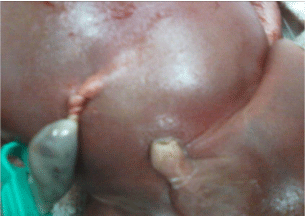
Figure 1
Features of the umbilical cord of the demised Twin 1.
Case Presentation
A 24-years-old, primigravida woman with a spontaneous MCDA twin pregnancy was for the first time admitted in our Division at 27 weeks for threatened preterm labour and Twin 1 fetal demise. Until this time, pregnancy was uncomplicated and the ultrasound examinations routinely performed, as well as the fetal echocardiograms, was normal. Laboratory evaluation was normal, with negative cervical and urine cultures, negative results for human immunodeficiency virus I, hepatitis B and C. TORCH testing and Parvovirus B19 assessment did not highlight ongoing infections. The woman was Rhesus negative, so the Kleihauer test was performed and anti-D immunoglobulins were administered. The ultrasound examination at then admission showed fetal hydrops (ascites, hydrothorax, nuchal edema) in the demised Twin 1 with normal growth, amniotic fluid pocket and Doppler in Twin 2. The inter-twin discordance estimated fetal weight was 4% with only mild difference in the amniotic fluid amount (vertical pool depth: Twin 1: 5.3 cm, Twin 2: 8.5 cm). All these findings were suggestive of an acute TTTS as a possible cause of the single-twin demise. Three days after the admission, an urgent Caesarean section was performed for an abnormal fetal non-stress test in Twin 2. After delivery, the cord of Twin 1 showed multiple torsions and a complete constriction of the cord localized at 2 cm from the fetal insertion side (Figure 1). The post-mortem examination of the dead twin revealed a weight of 800 g (percentile 10-50), a length of 44 cm and a head circumference of 30 cm. The autopsy didn’t show any structural congenital malformations. Placental histopathology showed a regular monochorial diamniotic twin placenta with no signs of inflammation or vascular malformations. The cord of twin 1 was 19 cm in length, with a normal vessels number that anyway appeared extremely thinned and almost empty. Placental and umbilical microbiologic examination was negative. The alive co-Twin (Apgar scores 3 and 7 at the 1st and 5th minute, respectively) weighed 980 g (percentile 50-90) and had no clinical or biochemical signs of cardiac failure or anaemia (central haemoglobin and haematocrit were 16.1 g/ dl and 49%, respectively). After birth, the baby developed respiratory distress requiring mechanical ventilation, and, successively, nasal CPAP. On the second day of life cerebral ultrasound examination highlighted moderate hyper echogenicities of the periventricular white matter and bilateral grade II intraventricular haemorrhage. At two months of life, signs of perinatal ischemic brain damage involving the white matter and the right post-rolandic cortex were shown by the brain MRI. At 5 months of life, the clinical follow-up showed normal growth but neurological evaluation highlighted mild hyper excitability and hypertonia.
Discussion
Twin monochorionic pregnancies are characterized by an increased incidence of both fetal and maternal complications and are at considerably higher risk of single-twin demise than are dichorionic twins [3]. This higher risk is mostly related to the placental architecture with vascular inter-twin anastomoses, which may predispose the twins to acute and chronic haemodynamic imbalance [4]. The TTTS occurs in 15% of monochorionic twin pregnancies and is one of the major risk factors for sIUFD. In our case, Twin 1 fetal demise was prenatally attributed to an acute TTTS according to the ultrasound findings of fetal hydrops in MCDA pregnancy without any evidence of inter-twin fetal weight discordance. However, the placental and cord macroscopic examination in the delivery suite documented severe cord torsion of the dead Twin 1. These observations suggested the hypothesis that fetal demise in Twin 1 may not be attributable to an acute TTTS but rather to the acute development of severe cord torsion. This hypothesis arisen from either the presence of the cord torsion in Twin 1, or from the absence of any signs and symptoms suggestive of TTTS in Twin 2. Umbilical Cord torsion (UCT) is a pathological accentuation of the normal helicoidally twisting of the cord, mostly localized at the fetal end [5], as in our case. The etiology of UCT is still poorly understood, but an increased cord length may represent one possible risk factor [5], as well as the congenital absence of the Wharton’s jelly [6]. A recently published series of 130 cases of UCT has highlighted that neither genetic factors nor the twinning are implicated in the etiology of UCT cases since the recurrence risk in subsequent pregnancies is low and the incidence of UCT in multiple gestations is similar to that reported in single ones [7]. The clinical signs of UCT can be different according to the speed development of the cord torsion: the slow occurrence may lead to chronic hypoxia with decreased blood flow, fetal growth retardation, and oligohydramnios, while its acute development may completely obstruct fetal-placental circulation, thereby causing heart failure, non-immune hydrops, and/or subsequent fetal death [8,9]. Although challenging, a proper prenatal ultrasound evaluation may be able to identify the presence of abnormal cord coiling: however, acute torsions are mostly unexpected and unpreventable [5]. Our observation is in keeping with the fact that sIUFD in MCDA pregnancy can be due to both unequal placental sharing, leading to TTTS, but also to abnormalities in the insertion, length or increased coiling of the umbilical cord. The present case report underlines the importance to consider and rule out all the differential diagnosis behind a sIUFD in MCDA pregnancy. Therefore, a carefully performed examination of the placenta and of the cords is essential to correctly investigate all the possible causes of sIUFD. The unexplained stillbirth rate is reported to be almost 30% but, hopefully, a full assessment (post-mortem analysis and placental investigation) might reduce it to a 5% rate [10].
Acknowledgement
We really would like to thank the guardians for giving permission to submit this case report, which will undoubtedly develop our understanding about this condition.
References
- Shek NW, Hillman SC, Kilby MD. Single-twin demise: pregnancy outcome. Best Pract Res Clin Obstet Gynaecol. 2014;28(2):249-63.
- Ong SS, Zamora J, Khan KS, Kilby MD. Prognosis for the co-twin following single-twin death: a systematic review. BJOG. 2006;113(9):992-8.
- Glinianaia SV, Obeysekera MA, Sturgiss S, Bell R. Stillbirth and neonatal mortality in monochorionic and dichorionic twins: a population-based study. Hum Reprod. 2011;26(9):2549-57.
- Mahony R, Mulcahy C, McAuliffe F, Herlihy CO, Carroll S, Foley ME. Fetal death in twins. Acta Obstet Gynecol Scand. 2011;90(11):1274-80.
- Pinar H, Carpenter M. Placenta and umbilical cord abnormalities seen with stillbirth. Clin Obstet Gynecol. 2010;53(3):656-72.
- Hallak M, Pryde PG, Qureshi F, Johnson MP, Jacques SM, Evans MI. Constriction of the umbilical cord leading to fetal death. A report of three cases. J Reprod Med. 1994;39(7):561-5.
- Rodriguez JI, Marino-Enriquez A, Suarez-Aguado J, Lapunzina P. Umbilical cord stricture is not a genetic anomaly: a study in twins. Pediatr Dev Pathol. 2008;11(5):363-9.
- Ben-Arie A, Weissman A, Steinberg Y, Levy R, Hagay Z. Oligohydramnios, intrauterine growth retardation and fetal death due to umbilical cord torsion. Arch Gynecol Obstet. 1995;256(3):159-61.
- Fleisch MC, Hoehn T. Intrauterine fetal death after multiple umbilical cord torsion-complication of a twin pregnancy following assisted reproduction. J Assist Reprod Genet. 2008;25(6):277-9.
- Flenady V, Koopmans L, Middleton P, Froen JF, Smith GC, Gibbons K, et al. Major risk factors for stillbirth in high-income countries: a systematic review and meta-analysis. Lancet. 2011;377(9774):1331-40.