Case Report
A Giant Phyllodes Tumor of the Breast: A Case Report in Pregnancy
Maria Grazia Tortoriello1, Rocco Cerra1, Maurizio Di Bonito2, Gerardo Botti2, Fabiola Gilda Cordaro3, Emilia Caputo3* and Raffaele Tortoriello1*
1Department of Senology, Istituto Nazionale Tumori Fondazione G. Pascale, Italy
2Anatomia Patologicae Citologica, Istituto Nazionale Tumori Fondazione G. Pascale, Italy
3Institute of Genetics and Biophysics–I.G.B., Italy
*Corresponding author: Caputo E and Tortoriello R, Department of Senology, Istituto Nazionale Tumori Fondazione G. Pascale, Naples, 80131- Italy
Published: 21 Mar, 2017
Cite this article as: Tortoriello MG, Cerra R, Di Bonito M,
Botti G, Cordaro FG, Caputo E, et al.
A Giant Phyllodes Tumor of the Breast:
A Case Report in Pregnancy. Ann Clin
Case Rep. 2017; 2: 1311.
ISSN: 2474-1655.
Abstract
Phyllodes Tumors (PTs) of the breast are rare biphasic fibro epithelial tumor. They account for less than 1% of primary breast tumors. They typically grow rapidly and clinically appears as breast lump in woman within a median age of 45 years. PTs are characterized by an enhanced intra canicular structure with leaf-like project ions into dilated lumens. In the 70% of cases, these tumors constitute benign unilateral lesions of the female breast. Less common are the malignant phyllodes, characterized by stromal pleomorphism and over growth, frequent mitosis and infiltrative borders. PTs, rarely, occur in pregnancy. Here, we present a case of 37 years old women with a giant malignant PT in her left breast, with a maximum diameter of about 24 cm, the largest reported in literature. It grew over 7months, during pregnancy. The histological examination of the resected tumor specimen, after its wide excision, revealed predominance of stromal hyper cellularity with a few altered epithelial component and a focal area showing a leaf-like pattern, consisting with a large malignant phyllodes tumor.
Introduction
Phyllodes tumors (PTs) of the breast are a rare and complex group of biphasic fibro-epithelial
neoplasms [1]. They represent less than 1% of primary breast neoplasms. PTs usually present
as breast lump and they are diagnosed in all age groups with a median age of presentation of 45
years. Rarely are diagnosed during pregnancy [2,3]. These tumors are often clinically benign and
are characterized by a rapid growth, with an increased intra canicular growth pattern with leaflike
projections. Malignant PTs are rare and exhibit an enhanced stromal pleomorphism and over
growth, frequent mitosis and in filtrative borders. Currently, biopsy is the too lused for diagnosing
phyllodes tumors. The treatment is surgery, by wide local excision with sufficient margin of normal
breast tissue or mast ectomy.
Here,we presented a case of a 37 years old women with a large breast left mass of about 24 cm
diameter. The mass was surgically removed by wide local excision and the histological examination
demonstrated the presence of a biphasic fibro epithelial neoplasm. The stromal component exhibited
an enhanced intra canalicular pattern and leaf like projections, accompanied by hyper cellularity
and moderate nuclear atypia. Stromal over growth was observed. The epithelial component also
showed alterations, consisting of marked adenosis are as, accompanied sometimes in lactation like
epithelial changes. Furthermore, areas of malignant degeneration were detected. All together these
changes were consistent with a giant malignant phyllodes tumor.
Case Presentation
A 37 years old Italian women presented in our hospital, in the first three month period of
pregnancy, with a small lump on the left breast above the nipple, measuring about 1.2 cm. Her only
significant past medical history was adenoids. She referred to have a miscarriage eat 36 years old,
and to be under hormonal therapy. She never smoked. She had thrombophilia and she was under
anticoagulant medication. She was an allergic individual to antibiotics, belonging to the cephalosporin
and penicillin class. She had not family history of breast or ovarian cancer. Ultra sound examination
demonstrated an odular formation on the left breast contained liquid, consistent with a suspicious
BI-RADSU4 tumor, according to the BI-RADS classification [4,5]. Successive examination, by coreneedle
biopsy, revealed a biphasic fibro epithelial neoplasm. The epithelial components hawed areas of slight iperplasia, while the stromal one presented spindle cells with
moderate hyper cellularity, leaf-like projections, without atypia and
mitosis, suggesting a doubtful benign phyllodes tumor. After she gave
birth, she came back to our hospital. She was breast feeding. At that
time, physical examination showed a massively enlarged left breast
mass, measuring about 24 cm in maximum diameter (Figure 1). She
stated that it was not painful. In particular, mammography revealed
that the left mammary gland body was entirely replaced from a
poly lobed nodular growth. The patient right breast and the rest of
clinical examination were normal. Examination by ultrasound breast
with repere revealed a large heterogeneous solid mass with internal
vascularity, replacing all normal left breast tissue. No suspicious
findings in the left lymph node as well asin the right breast and in the
right axilla were detected.
Further examination with a chest and abdomen ultra sound was
performed, which showed no evidence of metastatic lesion. The patient
under went to a wide local excision with sufficient surgical margin
resection (Figure 2A). The resected tumor specimen measured 24 x16
x4 cm (Figure 2B). Histological examination demonstrated a fibro
epithelial architecture with an exaggerated intra canalicular pattern
(Figure 3A) with leaf-like fronds (Figure 3B), accompanied by stromal
hyper cellularity (Figure 3C), and moderate nuclear atypia (Figure
3D). Alterations of the epithelial component were demonstrated,
consisting of marked adenosis areas, accompanied sometimes in
lactation-like epithelial changes. Stromal overgrowth (defined as the
presence of stroma without epithelium in at least one low-power field
as observed with a × 4 microscope objective) was observed. Amitotic activity of 11/10high-power fields (HPFs) was measured in an area
of stromal hyper cellularity. The tumors howed well defined borders.
The histological examination was consistent with a giant phyllodes
tumor, showing area of malignant degeneration.
Figure 1

Figure 1
Enlarged patient’s left breast.
Figure 2
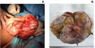
Figure 2
(A) Operative findings: wide local excision (B) The excised tumor
mass, 24 cm x 16 cm x 4 cm in size.
Figure 3
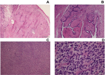
Figure 3
Histo pathological findings: histological analysis of the surgical
specimen demonstrated a diffuse fibro-epithelial lesion showing a few
elongated epithelium lined clefts with stromal mounds by low magnification
(A) A leaf-like pattern typical of phyllodes tumor was demonstrated at higher
magnification (B) Marked stromal hyper cellularity was observed (C) with
moderate nuclear atypia with alteration in nuclear size and irregular nuclear
membranes (D) All the tissue sections were stained by hematoxy line
osin.100 μm bar was reported.
Discussion
PTs of the breast are rare fibro epithelial neoplasms, which
can occur in all age groups, but predominantly occur in middle
aged women, with the average of 45 year old [6,7]. On clinically
examination, these tumors usually present as painless mass with an
average size of 4-5 cm [8,9]. They are classified into benign, border
line and malignant grade categories on the basis of their histological
properties, i.e. the degree of stromal cellularity and atypia, stromal
overgrowth, mitotic activity and the nature of their tumor borders
[1]. However, it is still difficult, not only, accurate and reproducible
grading classification of these tumors but also the discrimination
between benign phyllodes tumors and cellular fibro adenoma as
well as between the malignant phyllodes tumors and primary breasts
arcomaor spindle cell meta plastic carcinoma in all this spectrum of
the PTs. This is due to overlapping microscopic features among these
tumors. In addition to add more complexity to the PTs diagnosis
and categorization is their intra tumoral heterogeneity. It is in fact
no tun common that these tumors show benign lesions in some area
and characteristics of border line and malignant lesions in other areas
[10].
In our case, we observed a small lump (1.2 cm) on the left
breast consisting of a biphasic fibro epithelial neoplasm, in the first
trimester of pregnancy in 37 years Old Italian women. Only two cases
are reported in literature of phyllodes tumors diagnosed during the
first trimester of pregnancy [2,11]. Our case was not immediately
treated but after the patient gave the birth. She experienced rapid
growth during pregnancy, leading the left breast mass to a dimension
of about 24 cm, the largest phyllodes tumor reported in literature.
Rapid growth is one of the features of PTs and seven cases are
reported in literature with this property, suggesting that the growth
is more enhanced during pregnancy [12]. Further, this finding supports the idea that these tumors may be hormonally sensitive,
although this concept is highly debated [3]. In this case, our patient
referred a miscarriage at 36 years old, and to be under hormonal
therapy. Interesting, in 10 reported cases of PTs during pregnancy,
six presented a history of a previous breast mass [2,3,11,13-15]. Our
patient did not report any previous breast mass. Furthermore, she
had no family history of breast and ovarian cancer.
References
- LakhaniSR, Ellis IO, Schnitt SJ, Tan PH, vande Vijver MJ. World Health Organization Classification of Tumours of the Breast. Lyon. IARC. 2012;4:240.
- Blaker KM, Sahoo S, Schweichler MR, Chagpar AB. Malignant phylloides tumor in pregnancy. Am Surg. 2010;76(3):302-5.
- Sharma JB, Wadhwa L, Malhotra M, Arora R, Singh S. A case of huge enlargement of cystosarcoma phylloides of breast in pregnancy. Eur J Obstet Gynecol Reprod Biol. 2004;115(2):237-9.
- Mendelson EB, Baum JK, Berg WA, Merritt CR, Rubin E, BI-RADS: Ultrasound. In Breast Imaging Reporting and Data System: ACRBIRADS- Breast Imaging Atlas. American College of Radiology; 2002.
- Hille H, Vetter M, Hackelöer BJ. The accuracy of BI-RADS classification of breast ultrasound as a first-line imaging method. Ultraschall Med. 2012;33(2):160-3.
- Barrio AV, Clark BD, Goldberg JI, Hoque LW, Bernik SF, Flynn LW, et al. Clinicopathologic features and long-term outcomes of 293 phyllodes tumors of the breast. Ann Surg Oncol. 2007;14(10):2961-70.
- Karim RZ, Gerega SK, Yang YH, Spillane A, Carmalt H, Scolyer RA, et al. Phyllodes tumours of the breast: a clinico pathological analysis of 65 cases from a single institution. Breast. 2009;18(3):165-70.
- Bellocq JIARC. 2003:100-103.
- Rosen P. Fibro ephitelial Neoplasms. Lippincott-Raven. 1997:155-173.
- Tan BY, Acs G, Apple SK, Badve S, Bleiweiss IJ, Brogi E, et al. Phyllodes tumours of the breast: a consensus review. Histopathology. 2016;68(1):5- 21.
- Gentile LF, Gaillard WF, Wallace JA, Spigue LR, Alizadeh L, Lentz A, et al. A Case of a Giant Borderline Phyllodes Tumor Early in Pregnancy Treated with Mastectomy and Immediate Breast Reconstruction. Breast J. 2016;22(6):683-687.
- Belkacémi Y, Bousquet G, Marsiglia H, Ray-Coquard I, Magné N, Malard Y, et al. Phyllodes tumor of the breast. Int J Radiat Oncol Biol Phys. 2008;70(2):492-500.
- Simpson SA, Redstone J, Aziz MS, Bernik SF. Large malignant phyllodes tumor with rapid growth during pregnancy: images of a case. Breast J. 2007;13(6):620-1.
- Ray S, Basak S, Das S, Pal M, Konar H. Malignant phylloides tumor of breast in a pregnant woman with coincidental nulliparous vaginal prolapse. Iran J Med Sci. 2011;36(4):315-7.
- Pacchiarotti A, Frati P, Caserta D, Pacchiarotti A, Frega A, Moscarini M. First case of transformation for breast fibroadenoma to high-grade malignant cystosarcoma in an in vitro fertilization patient. Fertil Steril. 2011;96(5):1126-7.