Commentary
Axillary Mass
Mladen Mimica* and Danijel Pravdic
Department for Internal Diseases, Clinical Hospital Mostar, Bosnia and Herzegovina
*Corresponding author: Mladen Mimica, Department for Internal Diseases, Clinical Hospital Mostar, Bijeli Brijeg BB, 88000 Mostar, Bosnia and Herzegovina
Published: 13 Mar, 2017
Cite this article as: Mimica M, Pravdic D. Axillary Mass.
Ann Clin Case Rep. 2017; 2: 1299.
Commentary
Question
What is the most likely diagnosis in this asymptomatic 34-year-old woman, with painless left
axillary mass that became apparent in the advanced pregnancy (Figure 1 - arrow)?
• Sarcoidosis
• Hydradenitis suppurativa
• Abscess
• Galactocela
• Breast cancer metastasis
Answer: Galactocela
Explanation
A 34-year-old woman presented at the University Hospital with painless swelling of her
left axilla occurred during last phase of second pregnancy, with tendency for rapid growth after
childbirth, during lactation (Figure 1). There were no axillary’s mass during her first pregnancy.
General physical examination was unremarkable, without signs of local or systemic infection.
The mass became apparent at the last month of her second pregnancy. Finally axillary mass was
measuring 3x2 cm. Formation was solitary, soft, oval-shaped, mobile and painless, with normal
overlying skin. Mass has fluctuated, and content was white blurred. All routine hematological and
biochemical parameters, including leukocytes and C-reactive protein were normal.
Ultrasonography of left axillary mass showed an oval shape just below the skin with anechoic
blurred content surrounded by heterogeneous area which has the properties of glandular mammary
tissue (Figure 2). In some locations mass had well, and in other ones it had irregulary defined
margins. The finding was most consistent with galactocoele. After aspiration of the mass the milk
was drained, and definitive diagnosis of galactocele arising in axillary accessory breast tissue was
made. Periodic monitoring and, if necessary, drainage was advised.
An accessory breast gland is most commonly located in the axilla and may enlarge during the terminal phase of pregnancy and lactation. The accessory breast
glands mainly occur during the first pregnancy, and usually recur
in subsequent ones. Sarcoidosis is multi-system disease. Patients are
usually not asymptomatic and axillary lymphadenopathy is usually
bilateral. Hidradenitis suppurativa is chronic skin infection, and
tends to start after puberty. It is characterized by clusters of painful
abscesses, epidermoid cysts, sebaceous cysts and pilonidal cysts that
most commonly affects apocrine sweat gland bearing areas, such as
the underarms, often lead to scarring. The process is usually bilateral.
Abscess is accompanied by symptoms and signs of inflammation.
Breast cancer metastases are usually rounded, not oval, and
commonly associated with pathological changes in the corresponding
breast. Awareness of the variety of disease entities and characteristic
sonographic findings with ultrasound guided puncture can aid in
correct diagnosis of an axillary mass. A painless lump developing
during or a few weeks after ended breastfeeding is generally thought
to be a galactocele.
Figure 1
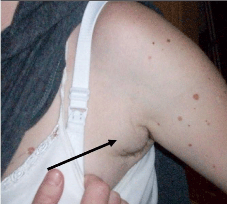
Figure 1
Painless swelling of her left axilla occurred during last phase of second pregnancy, with tendency for
rapid growth after childbirth, during lactation.
Figure 2
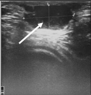
Figure 1
Ultrasonography of left axillary mass showed an oval shape just
below the skin with anechoic blurred content surrounded by heterogeneous
area which has the properties of glandular mammary tissue.