Clinical Image
Phytophotodermatitis
Dale Maharaj* and Fawwaz Mohammed
Department of Surgery, Caribbean Vein and Vascular Clinic St. Clair Hospital, Trinidad and Tobago
*Corresponding author: Dale Maharaj, Department of Surgery, Caribbean Vein and Vascular Clinic St. Clair Hospital, #18 Elizabeth Street St Clair, Port of Spain, Trinidad and Tobago
Published: 21 Feb, 2017
Cite this article as: Maharaj D, Mohammed F. Phytophotodermatitis. Ann Clin Case Rep. 2017; 2: 1278.
Clinical Image
This patient is a 65 year old Caucasian female who presented with a Hyperemic, desquamating rash on her hands. The rash was confined more to the dorsum of the hands with a burning sensation and areas of blistering. She noticed the rash a 2 days after squeezing limes and then being exposed to sunlight. In consultation with dermatologist she was diagnosed a Photodermatitis. Phytophotodermatitis is one of the 4 cutaneous dermatitis seen in humans. The ingredients needed to produce phytophotodermatitis include temporal exposure to both a photosensitizing substance, such as psoralens, and ultraviolet radiation. The primary skin lesion of phytophotodermatitis may range from delayed erythema (24-48 h) to frank blisters. The patient was seen in consultation with a dermatologist and counselled on avoiding future contact with offending agent (limes). The rash resolved in 3 days without intervention except for symptomatic relief. Topical Steroids, cool wet compresses and NSAIDS are usual treatment options.
Figure 1
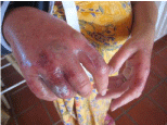
Figure 1
Picture of right hand showing desquamation.
Figure 2
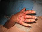
Figure 2
Right hand showing lesions.
Figure 3
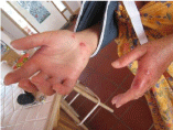
Figure 3
Palmar aspect Right hand showing improvement after treatment.