Clinical Image
Tuberculous Frontal Bone Osteomyelitis
B Mehdi, A Berriche*, L Ammari, R Abdelmalek, B Kilani and H Tiouiri Benaissa
Department of Infectious Diseases, Rabta Hospital, Tunisia
*Corresponding author: Aida Berriche, Department of Infectious Diseases, Rabta Hospital, Tunis, Tunisia
Published: 17 Feb, 2017
Cite this article as: B Mehdi, A Berriche, L Ammari,
R Abdelmalek, B Kilani, H Tiouiri
Benaissa. Tuberculous Frontal Bone
Osteomyelitis. Ann Clin Case Rep.
2017; 2: 1274.
Introduction
Tuberculosis is a major public health problem in endemic countries. It poses a diagnostic problem especially when the location is rare, such as the cranial vault. Tuberculous osteomyelitis of the skull accounts for 0.2 to 1.37% of all bone tuberculosis lesions. We report a case of a patient with tuberculous frontal bone osteomyelitis.
Clinical Image
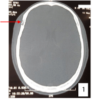
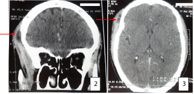
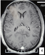
A 39 years old man presented with one month history of headache, nausea and vomiting, associated with night sweats without fever or impaired general condition. His past medical history was unremarkable. On admission, the patient was a febrile. A physical examination demonstrated a right frontal bulge which was painful, associated to fluctuation without inflammatory reaction around. A computed tomography (CT) of the brain showed a skin collection (22×5 mm) with osteolytic frontal lesion (Figure 1-3). The tuberculin skin test was positive (12 mm). Laboratory test results, including the complete blood profile and basic metabolic profile, were normal. The chest radiograph was normal. A sputum search of acid-fast bacilli was negative. A neurosurgery was performed: discharge of the abscess with frontal bone biopsy. Histological examination revealed an inflammatory process with epithelioïd granuloma and giant cell associated to a central caseating. The bacteriological examination was not performed. The patient was treated according to the specific sheme for treating bone tuberculosis, as recommended by our authoritis for 12 months. There was a decrease in symptoms with clinical improvement. The cerebral MRI, performed at 11 month of treatment, was normal (Figure 4). The patient is asymptomatic 3 years after the completion of treatment.
Figure 1
Figure 2 and 3
Figure 4
Discussion
Annually, thirty million active cases of tuberculosis worldwide and ten million new cases are recorded. The first case of tuberculous osteomyelitis was described in 1842. Tuberculous osteomyelitis is infrequent, and account for 0.2 to 1.37% of all bone lesions. Fifty percent of tuberculous osteomyelitis is seen in children less than ten years and, 71.42% in patients younger than 20 years [1]. The feature of our case is the patient's age (39 years). Mycobacterium tuberculosis spread in the skull through blood from a primary latent focus. Then, it attaches in bone tissue causing destruction and abscess with or without fistula [2]. The dura is an effective barrier to the contiguous spread of all infections [3]. The forms associated with tuberculoma, encephalitis and tuberculous meningitis are exceptional [2]. The diagnosis should be considered in any abscess or chronic suppuration [4,5]. Positive tuberculin skin test and the existence of another tuberculosis location are diagnosis elements [1]. Diagnosis is based on histology, showing an inflammatory process with epithelioid granuloma and giant cell associated to a central caseating, with or without isolation of Mycobacterium tuberculosis. Medical treatment prescribed for a period of 9 to 18 months associated to surgical treatment is mandatory for good outcome [6].
Conclusion
Tuberculous osteomyelitis is infrequent because of low bone blood supply. It preferentially affects the frontal and parietal areas. Diagnosis should be considered in any chronic scalp suppuration. Favorable outcome needs good adherence to medication.
References
- Van Dellen A, Nadvi SS, Nathoo N, Ramdial PK. Intracranial tuberculosis subdural empyema: case report. Neurosurgery. 1998; 43: 370-373.
- Mishra SK, Nigam P. Tuberculosis of flat bones. Indian J Chest Dis Allied Sci. 1984; 26: 174-176.
- M Boubrik, S Ait Benalib. Tuberculous osteitis of the skull vault: in two cases. Pediatric Archives. 2011; 18: 397-400.
- Barton CJ. Tuberculosis of the vault of the skull. Br J Radiol. 1961; 34: 286-290.
- Rudman IE. Tuberculosis of the bones of the skull. Med Times. 1965; 93: 910-913.
- Jadhav RN, Palande DA. Calvarial tuberculosis. Neurosurgery. 1999; 45: 1345-1350.


