Clinical Image
Large Esophageal Diverticula Concomitant Achalasia Treated by POEM
Huang Xiaoquan1,2 and Chen Shiyao1,2*
1Endoscopy Center and Endoscopy Research Institute, Fudan University, China
2Department of Gastroenterology, Fudan University, China
*Corresponding author: Chen Shiyao, Endoscopy Center and Endoscopy Research Institute, Department of Gastroenterology, Zhongshan Hospital, Fudan University, 180 Fenglin Road, Shanghai, 0086200032, China
Published: 14 Feb, 2017
Cite this article as: Xiaoquan H, Shiyao C. Large
Esophageal Diverticula Concomitant
Achalasia Treated by POEM. Ann Clin
Case Rep. 2017; 2: 1270.
Clinical Image
An 85-year old presented with severe pneumonia and reflux esophagitis. He had recently progressively vomit and dysphagia for about ten months. Barium contrast esophagography showed large esophageal diverticulum concomitant achalasia (Figure 1A). After drug treatment and esophageal stent implantation for two months. He switched to catheterized nasal feeding tube, and he was able to receive endoscopic treatment. A gastrografin-contrast study followed by computed tomographic scan were confirmed before endoscopic treatment (Figure 1B and C). He was successfully managed by peroral endoscopic myotomy (POEM) and endoscopic operation for both achalasia and large esophageal diverticulum (Figure 1D). He underwent oesophageal gastrografincontrast study soon after the procedure, which verified no perforation and smooth passage through the gastroesophageal junction. His symptoms abated, and he was discharged after oral refeeding on the ninth day of hospitalization.
Figure 1
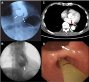
Figure 1
(A) Barium contrast esophagography showed large esophageal diverticulum concomitant achalasia.
(B and C) Gastrografin-contrast study followed by computed tomographic scan. (D) Endoscopic operation for
both achalasia and large esophageal diverticulum.