Case Report
Tension Pneumocephalus as a Complication of Intracranial Pressure Monitoring
Jonathan A Heaney* and YC Peter Gan
Department of Neurosurgery, Waikato Hospital, New Zealand
*Corresponding author: Jonathan A Heaney, Department of Neurosurgery, Waikato Hospital, Pembroke Street, Hamilton, New Zealand
Published: 09 Feb, 2017
Cite this article as: Heaney JA, Peter Gan YC. Tension
Pneumocephalus as a Complication of
Intracranial Pressure Monitoring. Ann
Clin Case Rep. 2017; 2: 1266.
Abstract
We present the case of tension pneumocephalus resulting as a complication of intracranial pressure monitoring in a non trauma patient. Tension pneumocephalus is a neurosurgical emergency requiring early recognition and management. Our patient’s CT head showed the typical Mount Fuji sign which prompted urgent neurosurgical decompression with favourable results. This manuscript also includes a description of the diagnosis and pathophysiology of pneumocephalus. This is the first published case to describe tension pneumocephalus resulting from intracranial pressure monitoring in a non trauma patient.
Keywords: Tension pneumocephalus; Mt Fuji sign; Intracranial pressure monitoring
Introduction
Pneumocephalus is a term used to describe the presence of intracranial air. Common causes
include trauma, infection, cerebrospinal fluid (CSF) leak, and any fistula or conduit between
the intracranial cavity and external air. Pneumocephalus has been reported to occur in all cases
following supratentorial craniotomy [1]. Areas of extreme hypodensity (Hounsfield coefficient
-1000) in the subdural space on computed tomography (CT) is characteristic of pneumocephalus.
If the volume of pneumocephalus is sufficient enough to exert mass effect on the brain, it is termed
tension pneumocephalus and considered a neurosurgical emergency requiring decompression
before brain herniation and death result.
Intracranial pressure (ICP) monitoring is commonly performed and may be achieved with the
placement of either subdural, extradural, intraparenchymalor intraventricular devices. Although a
relatively safe procedure, complications may include infection, haemorrhage and malposition. We
present a case of tension pneumocephalus as a complication of ICP monitoring.
Case Presentation
A 61 year-old gentleman was electively admitted for intracranial pressure monitoring. Five years prior a right-sided ventriculo-peritoneal shunt (STRATA II valve) had been inserted for post-traumatic delayed hydrocephalus. He had progressive worsening of headaches since shunt insertion, despite the shunt being set at the lowest possible setting of 0.5. The characteristics of his headaches were unclear but suggested shunt malfunction. The CT head showed dilated ventricles with no periventricular lucency (Figure 1). He thus presented for intracranial pressure monitoring. Under general anaesthesia an intraparenchymal pressure monitor (Codman Micro sensor Basic Kit bolt) was inserted at Kocher’s point on the right side via twist drill craniostomy. Opening pressure was measured at 10 mmHg and the patient awoke from anaesthesia unremarkably. The ICP readings were less than 10 mmHg throughout whilst the ICP monitor was in situ. Overnight the monitor and bolt became dislodged. The bolt and monitor were removed and the wound closed with sutures. Over the next hour the patient became increasingly restless and agitated, began vomiting and his GCS decreased from 15 to 12 (M6, E4, V2). A CT demonstrated classical tension pneumocephalus with Mt. Fuji sign and air within the cisterns (‘air bubble sign’) (Figure 2A and B). He underwent urgent decompression through bilateral frontal burr holes. He had an immediate and full recovery to his pre-operative state and was discharged two days later.
Figure 1
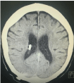
Figure 1
Axial CT demonstrating adequate placement of ventriculoperitonel
shunt and ventriculomegaly.
Figure 2
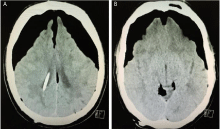
Figure 2
(A,B) Axial CT showing Mount Fuji sign and air in basal cisterns.
Note seperation and depression of frontal lobes.
Discussion
Tension pneumocephalus results from the one-way movement and expansion of air into the
subdural space. This one-way movement has been attributed to two possible mechanisms. The first,
known as the ‘inverted pop bottle’ mechanism, was described by Lunsford et al in 1979 [2]. By this mechanism, as CSF leaks, air is drawn into the cranium akin to
air bubbles rising in an inverted soda bottle as fluid pours out. The
second, known as the ‘ball-valve’ mechanism describes how once air
is trapped intracranially it cannot escape through the channel whence
it came. This was the most likely mechanism in our case, with the ICP
monitor insertion site being the one way valve.
The Mount Fuji and air bubble signs are pathognomonic of
tension pneumocephalus [3] and result from air pressure exerted
on the brain leading to depression and separation of the frontal
lobes, which may ultimately manifest as brain herniation and death.
Tension pneumocephalus as a complication of intracranial pressure
monitoring was first described by Vitali et al. [4] in a trauma patient
who had a subdural cup catheter ICP monitor inserted. To the best
of the authors knowledge, this is the first published case of tension
pneumocephalus occurring in a patient electively admitted for ICP
monitoring using an intraparenchymal pressure monitor. Our patient
may have been at increased risk of air ingress given he had a VP shunt
in situ which will have augmented any pressure gradient between the
atmosphere and intracranial space. Indeed, tension pneumocephalus
is recognised as a rare complication of VP shunt insertion surgery [5].
Conclusion
Intracranial pressure monitoring is commonly performed in both trauma and elective neurosurgical patients. This rare complication demonstrates upmost care must be employed when securing the monitor and during scalp closure to prevent this potentially fatal complication.
References
- Reasoner DK, Todd MM, Scaman FL. The incidence of pneumocephalus after supratentorial craniotomy. Observations on the disappearance of intracranial air. Anaesthesiology. 1994; 80: 1008-1012.
- Lunsford LD, Maroon JC, Sheptak PE, Albin MS. Subdural tension pneumocephalus. Report of two cases. J Neurosurg. 1979; 50: 525-527.
- Ishiwata Y, Fujitsu K, Sekino T, Fujino H, Kubokura T, Tsubone K, et al. Subdural pneumocephalus following surgery for chronic subdural haematoma. J Neurosurg. 1988; 68: 58-61.
- Vitali A, Roux A. Tension pneumocephalus as a complication of intracranial pressure monitoring: A case report. Ind J Neurotrauma. 2007; 4: 115-118.
- Ferrante L, Santoro A, Mastronardi L, Acqui M. Tension pneumocephalus after CSF shunting procedures. Br J Neurosurg.1988; 2: 269-272.