Clinical Image
A Clinical Diagnosis of Tuberous Sclerosis
Alani A* and Ahmad K
Department of Dermatology, University Hospital Limerick, Ireland
*Corresponding author: Angela Alani, Department of Dermatology, University Hospital Limerick, Limerick, Ireland
Published: 30 Jan, 2017
Cite this article as: Alani A, Ahmad K. A Clinical Diagnosis
of Tuberous Sclerosis. Ann Clin Case
Rep. 2017; 2: 1244.
Clinical Image
A 26 year-old lady presented for evaluation of an enlarging purple nodule on her left cheek as it bled on few occasions. She had a history of seizures and learning disability but no relevant family history. Examination revealed a large purple nodule measuring 47x39 mm on the left cheek (Panels A and B) (Figure 1) consistent with cutaneous angiofibroma. There were numerous adenoma sebaceum on her face with a hypomelanotic macule and multiple large Café au lait patches on her trunk and periungal fibromas on her fingers and toes. Excisional biopsy of facial nodule was consistent with angiofibroma. A clinical diagnosis of tuberous sclerosis (TS) was made. She was referred to neurology and had a normal Magnetic Resonance Imaging of brain. TS is an autosomal dominant disorder, affects multiple organs including brain, skin, eyes and kidneys. Up to 70% are thought to have new mutation of two different genes TSC1 and TSC2.
Figure 1
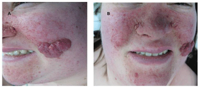
Figure 1
Large purple nodule with cutaneous angiofibroma.