Clinical Image
Abdominal Trompe-l’oeil Spirals
Presotto F*, Fascì-Spurio F, Serafini F and Zancanaro A
Unit of Internal Medicine, Hospital dell’Angelo, Italy
*Corresponding author: Fabio Presotto, Unit of Internal Medicine, Hospital dell’Angelo, Mestre- Venice, Italy
Published: 13 Jan, 2017
Cite this article as: Presotto F, Fascì-Spurio F, Serafini F,
Zancanaro A. Abdominal Trompe-l’oeil
Spirals. Ann Clin Case Rep. 2017; 2:
1234.
Clinical Image
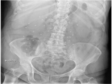
A 69-year-old woman arrived to our Department of Medicine for abdominal pain, hyperpyrexia and acute confusional state. Patient’s history was remarkable for bipolar syndrome, severe binge eating disorder with morbid obesity. In 2009 she underwent to bariatric surgery followed by 57 kg weight lost, from initial 142 kg. Height was 160 cm. Because of a wide laparocele, in 2010 she underwent to unspecified surgical repair. She was daily taking valproic acid 1.000 mg fluoxetine 20 mg, and alprazolam 0.50 mg. Physical examination showed diffuse abdominal tenderness, high fever, and microhematuria. The patient was not collaborating and neurological evaluation revealed somnolence and confused state, conditions ascribed to fever (39.3°C) or likely to inappropriate assumption of psychoactive drugs. Her initial blood examination showed normal leukocytes (4.500 per micro L) with mild anemia (Hb 12 g/dL) and normal C-reactive protein. An emergent plain film of the abdomen in anteroposterior projection showed multiple radiopaque spiral foreign bodies (Figure 1).
Discussion
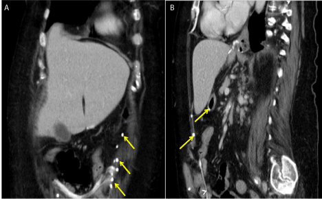
The initial suspicion was foreign metallic body’s ingestion (clothespins?) in a psychiatric patient with binge eating disorder. However, abdominal computed tomography described radiopaque devices consistent with a mesh and metallic spirals detectable in herniated bowels. No intestinal perforation or obstruction was found (Figure 2). It was then becoming clear that the foreign bodies were indeed spiral tacks employed for intraperitoneal mesh fixation by laparocele repair operation. Different fixation devices can be incidentally observed on plain abdominal radiographs. Materials from which mesh is manufactured are usually derived from polypropylene and functions as a bridge across defective tissues. The mesh is usually located in preperitoneal layers and held in place with tissue glue or metallic tacks [1]. Length of tacks is about 5 mm. They occasionally penetrate the neighboring structures, migrate into the peritoneal cavity and induce bowel perforation due to their sharp plug components [2,3]. If not carefully fixed tacks could fall into the abdominal cavity or the sharp end of a tacker not buried properly may cause injuries with movements. The patient had a final diagnosis of pyelonephritis and she was successfully treated with a course of intravenous ciprofloxacin.
Figure 1
Figure 1
Abdominal anteroposterior plain radiograph showing multiple spiral-shaped radiopaque metal bodies.
Figure 2
Figure 2
Abdominal coronal (a) and sagit cross sections showing a mesh in the abdotal (b) computed
tomography minal wall (white arrows) and multiple coiled radiopaque metal autosuture tacks (yellow arrows).
References
- Jamadar DA, Jacobson JA, Girish G, Balin J, Brandon CJ, Caoili EM, et al. Abdominal wall hernia mesh repair: sonography of mesh and common complications. J Ultrasound Med. 2008; 27:907-917.
- Haltmeier T, Groebli Y. Small bowel lesion due to spiral tacks after laparoscopic intraperitoneal onlay mesh repair for incisional hernia. International J Surgery Case Reports. 2013; 4: 283-285.
- Golash V. Large gut fistula due to a protruding spiral tacker after laparoscopic repair of a ventral hernia. Oman Medical Journal. 2008; 23: 50-52.


