Clinical Image
Bilateral Double-Layered Patella (DLP) with Suprapatellar Impingement
João Araújo*
Department of Radiology, Centro Hospitalar do Porto, Portugal
*Corresponding author: João Araújo, Department of Radiology, Centro Hospitalar do Porto, Portugal
Published: 10 Jan, 2017
Cite this article as: Araújo J. Bilateral Double-Layered
Patella (DLP) with Suprapatellar
Impingement. Ann Clin Case Rep.
2017; 2: 1231.
Clinical Image
A 22 year-old man presented complaining of anterior knee pain and no history of trauma.
A Magnetic Resonance Imaging (MRI) and Computed Tomography (CT) were performed,
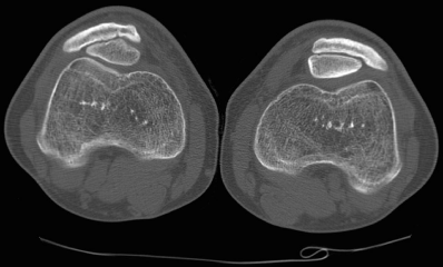
demonstrating two patellar layers bilateral, one anterior and one posterior on both knees, confirming
the diagnosis of bilateral DLP. We found the anterior osseous patellar layer attached to the extensor
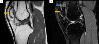
mechanism. In the MRI, both knees had fluid like signal replacing the normal supra patellar fat pad,
which we assumed to be the cause of the paindue to the impingement with the femoral trochlea. No
significant chondropathy was founded.
The DLP is a rare entity [1] that consists of two distinct patellar layers, one anterior and one
posterior. The literature admits frequent association of DLP with autosomal recessive form of multiple epiphyseal dysplasia (MED) [2,3]. An all body X-ray was performed and there were no
signs that suggest that association.
Figure 1
Figure 1
Axial bone window CT of the knees demonstrating two patellar layers and normal patella femoral joint
space.
Figure 2
Figure 2
Sagittal proton density fast spin echo MRI (A) and sagittal proton density with fat suppression fast spin
echo MRI of the left knee demonstrating cartilage tissue between the two layers (yellow arrow). The anterior
osseous patellar layer is attached to the extensor mechanism. There´s also fluid like signal replacing the normal
suprapatellarfat pad (blue arrow).
References
- Minh D Nguyen, Joshua S Everhart , Megan M May, David C Flanigan. Bilateral Double-Layered Patella: MRI Findings and Fusion with Multiple Headless Screws. J Bone Joint Surg. 2013; 3: e50.
- Rubenstein Joel D, Monique S Christakis. Fracture of Double Layered Patellain Multiple Epiphyseal Dysplasia: Radiology. Radiology. 2006; 239: 911-913.
- Ramachandran G, Mason D. Double-layered patella: marker for multiple epiphyseal dysplasia. American Journal Orthop (Belle Mead NJ). 2004; 33: 35-36.


