Clinical Image
What’s Your Diagnosis?
Jeonghyun Shin*
Department of Dermatology, Inha University, South Korea
*Corresponding author: Cheng-Ku Tsai, Department of Otolaryngology, Taichung Chi Hospital, No.11812F-6, Yuk Tak Road, North District, Taichung City, Taiwan, China
Published: 27 Dec, 2016
Cite this article as: Tsai C-K, Wu H-P. A Case of Nasal
Squamous Cell Carcinoma with the
Clinical Symptom of Recurrent Epistaxis
in a Case of Nasal Squamous Cell
Carcinoma. Ann Clin Case Rep. 2016;
1: 1224.
Clinical Image
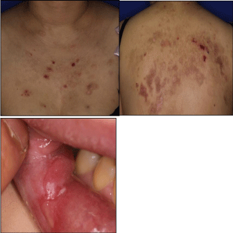
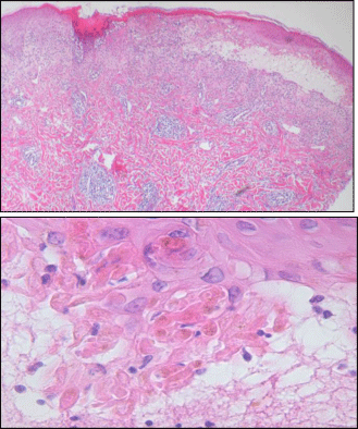
A 29-year-old woman visited to our department with presenting recurrent pruritic erythematous crusted patches for 4 years. She complained pruritic tense bullae initially developed. Physical examination showed scattered crusted patches with erosion on the back, chest, and forearms (Figure 1A and B). Buccal mucosa ulcerative lesion was also seen (Figure 1C). She has been followed up at the Rheumatology department for recurrent oral and genital ulcer with tentative diagnosis as Behcet disease. Her family history was not contributable. With laboratory examination, the level of peripheral blood circulating immune complex was increased to 3.52. Others were within normal limit. Skin biopsy was performed (Figure 2). Direct immunofluorescence study was negative.
Figure 1
Figure 1
Erythematous crusted patches and brownish pigmented patches on the chest and back (A & B).
Buccal mucosal ulcer was found (C).


