Clinical Image
Hepatic Portal Venous Gas
Boldizsar Kovacs* and Bernhard Magdeburg*
Regional Hospital of Wetzikon, Switzerland
*Corresponding author: Boldizsar Kovacs, Regional Hospital of Wetzikon, Spitalstrasse 66, 8620 Wetzikon, Switzerland
Published: 23 Dec, 2016
Cite this article as: Kovacs B, Magdeburg B. Hepatic Portal
Venous Gas. Ann Clin Case Rep. 2016;
1: 1221.
Clinical Image
A 75-year-old male with recurring adhesive ileus presented with abdominal pain. On clinical examination diminished bowel sounds were noted and the following abdominal x-ray confirmed our suspicion of intestinal obstruction. Following an exploratory laparotomy with extensive adhesiolysis and resection of necrotic bowel, septic shock rapidly developed and the patient was transferred to our ICU. A CT-scan was performed which revealed an extensive hepatic portal venous gas (HPVG) (Figure 1), confirmed by bedside abdominal ultrasound (Figure 2). The differentiation of HPVG from areobilia is made radiologically or sonographically. In the case of areobilia, gas is distributed centripetally due to the flow of bile towards the main biliary duct. The portal-venous flow on the other hand is centrifugal, thus gas flows peripherally. Therefore, when gas is spotted within 2cm of the liver capsule, the diagnosis of HPVG is more probable.
Figure 1
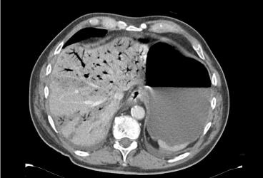
Figure 1
Axial abdominal CT scan showing hepatic portal venous gas.
Figure 2
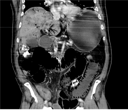
Figure 2
Coronal abdominal CT scan showing hepatic Portal venous gas and intestinal pneumatosis.