Case Report
18F-FDG PET/CT Imaging of Parotid Gland Sarcoidosis in a Young Scandinavian Male
Rikke Broholm*, Jens Bülow and Ali Asmar
Department of Clinical Physiology and Nuclear Medicine, Bispebjerg University Hospital, Denmark
*Corresponding author: Rikke Broholm, Department of Clinical Physiology and Nuclear Medicine, Bispebjerg University Hospital, Denmark
Published: 19 Dec, 2016
Cite this article as: Broholm R, Bülow J, Asmar A. 18F-FDG
PET/CT Imaging of Parotid Gland
Sarcoidosis in a Young Scandinavian
Male. Ann Clin Case Rep. 2016; 1:
1215.
Abstract
Sarcoidosis is a multisystem granulomatous disease of unknown cause that primarily involves the lungs. Extrapulmonary sarcoidosis is seen in more than 30% of patients. We report a case of a 29-year old man presenting with bilateral enlargement of the parotid glands combined with fever, discrete dyspnea, and fatigue. Histopathology from the parotid gland indicated sarcoidosis. An 18F-FDG PET/CT performed to visualize the organ extent demonstrated increased 18F-FDG uptake in the parotid glands and in bilateral mediastinal and hilar lymph nodes. The patient was diagnosed with sarcoidosis.
Introduction
Sarcoidosis is a multisystem granulomatous disorder of unknown cause that may involve any
organ. Pulmonary involvement predominates as well as affection of the intrathoracic lymph nodes,
eyes and skin; however it may affect any organ [1]. Extrathoracic manifestation is common and
seen in more than 30% of patients, typically in combination with thoracic involvement. Sarcoidosis
occurs at all ages, however with the incidence peaking at 20-39 years.
Systemic symptoms are weight loss, fatigue and night sweats but most patients present with
cough and dyspnea because of pulmonary involvement. The diagnosis of sarcoidosis is based on
clinical and radiological findings as well as histological findings of non-caseating epithelioid-cell
granulomas. Other causes of granulomas must be ruled out. Chest radiography and computed
tomography (CT) are used as routine diagnostic procedures in the evaluation of pulmonary
sarcoidosis but they are unable to detect active inflammation.
18F-fluorodeoxyglucose positron-emission tomography (18F-FDG PET)/CT has shown useful in
assessing the extent of organ involvement and in suggesting the organs that might be candidates
for diagnostic biopsy [2]. Furthermore, 18F-FDG PET/CT seems to contribute to a better evaluation
of the extrapulmonary involvement and it may identify occult granulomatous lesions that are not
detected by physical examination, conventional thoracic radiography or CT [3,4]. Additionally, the 18F-FDG PET/CT has been compared to 67Ga-scintigraphy in patients with sarcoidosis illustrating a
superior sensitivity, especially in depicting sites of extrathoracic involvement [5].
Here we report a case of sarcoidosis detected by 18F-FDG PET/CT.
Case Presentation
A 29-year old man with a history of bilateral enlargement of the parotid glands, alternating fever,
discrete dyspnea, and fatigue for several months was admitted to the hospital. Before admission, the
patient had been examined by an otorhinolaryngologist and a fine needle biopsy from the parotid
gland was conducted, demonstrating granulomas, indicative of sarcoidosis. The patient had felt
enlargement of the inguinal lymph nodes but had otherwise no other discomforts. Due to suspicion
of sarcoidosis, an 18F-FDG PET/CT was conducted to visualize the extent of organ involvement.
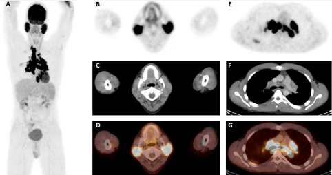
The 18F-FDG PET/CT revealed significantly increased 18F-FDG uptake in bilateral enlarged parotid
glands (Figure 1A and D) and in bilateral mediastinal and hilar lymph nodes, predominantly right paratracheal adenopathy (Figure 1E and G). No enlargement or increased 18F-FDG uptake was observed in the inguinal lymph nodes. Measurement of the serum angiotensin-converting-enzyme (ACE)
demonstrated highly elevated level (166 U/l) (normal upper limit < 115 U/l).
The 18F-FDG PET/CT findings, elevated level of serum ACE, and histopathological examination
of parotid gland biopsy with non-caseating epithelioid-cell granulomas confirmed the diagnosis of
sarcoidosis.
As the patient’s symptoms were regressing, no treatment was
initiated.
Figure 1
Figure 1
Whole-body 18F-FDG PET projection (A) and transaxial 18F-FDG PET/CT projections demonstrated diffuse high 18F-FDG distribution in bilateral enlarged
parotid glands (B-D) and in bilateral mediastinal and hilar lymph nodes (E-G).
Discussion
In 1990 Sulavik and colleagues described increased symmetrical
lacrimal gland and parotid gland 67Ga-citrate uptake combined
with normal accumulation of the radionuclide in the nasopharynx
(“panda” appearance) in 79% of sarcoidosis patients [6]. Furthermore,
a distinctive intrathoracic lymph node 67Ga -uptake pattern was
observed, resembling the Greek letter lambda (λ). The simultaneous
"lambda" and "panda" patterns were observed only in sarcoidosis
patients and this was considered highly specific for sarcoidosis [6].
Therefore, it has been argued that the combination of the "lambda"
and "panda" sign may obviate a histopathological examination.
Enlargement of the parotid glands is rarely seen in patients with
sarcoidosis (~6%) [7].
The appearance of hypermetabolic intrathoracic
lymphadenopathy detected by 18F-FDG PET/CT in patients with
sarcoidosis is comparable to the "lambda" sign on the gallium
scintigraphy as well as the bilateral involvement of the parotid and
lacrimal glands with high 18F-FDG uptake, resembling the "panda"
sign [8]. The typical "panda" appearance is however partially obscured
because of the high physiological 18F-FDG uptake of the brain. In our
patient the "lambda" and "panda" signs coexisted on the 18F-FDG
PET/CT although involvement of the lacrimal glands could not be
visualized.
18F-FDG PET/CT is increasingly used in the diagnostic workup
of pulmonary and mediastinal tumors that are suspected to be
malignant. However, sarcoid lesions can demonstrate high 18F-FDG
uptake mimicking malignant processes such as lymphoma or lymph
node metastases. Irrespective of the combination of the “lambda” and
“panda” signs detected by 18F-FDG PET, histological confirmation
should be mandatory.
In active sarcoidosis, a significant increased metabolism in
the active lesions can be detected by an increased 18F-FDG uptake.
However, after immunosuppressive therapy or spontaneous
regression, the metabolism of the lesions may decrease [4,9,10],
probably prior to morphological changes. Thus, 18F-FDG PET/CT may be a valuable adjunct to the clinical examination in monitoring the response to therapy.
Although the serum level of ACE is elevated in up to 60% of
sarcoidosis patients, it is never diagnostic since elevation can be seen
in other diseases [7]. It may however decrease after corticosteroid
treatment or spontaneous improvement [1]. In this patient no
treatment was initiated as spontaneous regression was observed.
Conclusion
18F-FDG PET/CT is valuable in demonstrating active lesions of sarcoidosis, both thoracic and extrathoracic involvement. Furthermore, 18F-FDG PET/CT might be useful in monitoring treatment response in patients with sarcoidosis; however, costs and radiation expose should always be taken into account.
References
- Iannuzzi MC, Rybicki BA, Teirstein AS. Sarcoidosis. N Engl J Med. 2007; 357: 2153-2165.
- Kruger S, Buck AK, Mottaghy FM, Pauls S, Schelzig H, Hombach V, et al. Use of integrated FDG-PET/CT in sarcoidosis. Clin Imaging. 2008; 32: 269-273.
- Aksoy SY, Ozdemir E, Senturk A, Seyda Türkölmez. A case of sarcoidosis diagnosed by positron emission tomography/computed tomography. Indian J Nucl Med. 2016; 31: 198-200.
- Teirstein AS, Machac J, Almeida O, Lu P, Padilla ML, Iannuzzi MC. Results of 188 whole-body fluorodeoxyglucose positron emission tomography scans in 137 patients with sarcoidosis. Chest. 2007; 132: 1949-1953.
- Nishiyama Y, Yamamoto Y, Fukunaga K, Takinami H, Iwado Y, Satoh K, et al. Comparative evaluation of 18F-FDG PET and 67Ga scintigraphy in patients with sarcoidosis. J Nucl Med. 2006; 47: 1571-1576.
- Sulavik SB, Spencer RP, Weed DA, Shapiro HR, Shiue ST, Castriotta RJ. Recognition of distinctive patterns of gallium-67 distribution in sarcoidosis. J Nucl Med. 1990; 31: 1909-1914.
- Statement on sarcoidosis. Joint Statement of the American Thoracic Society (ATS), the European Respiratory Society (ERS) and the World Association of Sarcoidosis and Other Granulomatous Disorders (WASOG) adopted by the ATS Board of Directors and by the ERS Executive Committee, February 1999. Am J Respir Crit Care Med. 1999; 160: 736-755.
- Oksuz MO, Werner MK, Aschoff P, Pfannenberg C. 18F-FDG PET/CT for the diagnosis of sarcoidosis in a patient with bilateral inflammatory involvement of the parotid and lacrimal glands (panda sign) and bilateral hilar and mediastinal lymphadenopathy (lambda sign). Eur J Nucl Med Mol Imaging. 2011; 38: 603.
- Braun JJ, Kessler R, Constantinesco A, Imperiale A. 18F-FDG PET/CT in sarcoidosis management: review and report of 20 cases. Eur J Nucl Med Mol Imaging. 2008; 35: 1537-1543.
- Treglia G, Annunziata S, Sobic-Saranovic D, Bertagna F, Caldarella C, Giovanella L, et al. The role of 18F-FDG-PET and PET/CT in patients with sarcoidosis: an updated evidence-based review. Acad Radiol. 2014; 21: 675-684.

