Case Report
Total Elbow Arthroplasty for Complete Bony Ankylosis of the Elbow in Patients with Rheumatoid Arthritis: A Report of Three Cases
Masanori Nakayama*, Yu Sakuma, Tetsuji Hosozawa, Hitoshi Imamura, Koichiro Yano and
Katsunori Ikari
Department of Orthopedic Surgery, Institute of Rheumatology, Tokyo Women's Medical University, Japan
*Corresponding author: Masanori Nakayama, Department of Orthopedic Surgery, Institute of Rheumatology, Tokyo Women's Medical University, 10-22 Kawada-cho, Shinjuku-ku, Tokyo, 162-0054, Japan
Published: 09 Dec, 2016
Cite this article as: Nakayama M, Sakuma Y, Hosozawa
T, Imamura H, Yano K, Ikari K. Total
Elbow Arthroplasty for Complete Bony
Ankylosis of the Elbow in Patients with
Rheumatoid Arthritis: A Report of Three
Cases. Ann Clin Case Rep. 2016; 1:
1213.
Abstract
We report three cases of Total Elbow Arthroplasty (TEA) for complete bony ankylosis in patients with Rheumatoid Arthritis (RA). Two cases had trauma around the elbow joint before ankylosis. At the most recent examinations, the average arc of motion was 85 degrees among patients. Two patients satisfied with surgical outcomes, however, one patient complained of residual pain and numbness. There were some reports about TEA of ankylosed elbow, however, there were few reports limited to ankylosed elbow with RA. TEA for ankylotic elbows in patients with RA is a good surgical option to restore adequate function. To gain a good outcome, it should be evaluated other joints in the extremity in order to properly evaluate the mobility and function of the elbow and to prepare a comprehensive plan of pre- and post-operative management that takes into consideration the full motion of the upper extremity.
Keywords: Ankylotic elbow; Rheumatoid arthritis, Total elbow arthroplasty
Introduction
Rheumatoid Arthritis (RA) is a common disease characterized by chronic synovitis and
resulting in joint destruction. Recent advancements in the diagnostic process and treatment of RA
have decreased joint surgery, including synovectomy or joint replacement, over the last decade [1]. Nevertheless, some patients with severe joint destruction occasionally develop severe contractures
or ankylosis which impair the Activities of Daily Living (ADL).
The incidence of elbow joint involvement in Rheumatoid Arthritis (RA) is approximately 50%
[2]. Destruction of the elbow joint seriously impairs upper limb function. Moreover, complete
ankylosis of the elbow results in severe disability and functional limitations, especially when other
joints in the ipsilateral upper extremity also have limited motion [3]. In a previous study not limited to RA patients, the analysis of compensatory movement after arthrodesis of the elbow showed
compromised ability, despite a significantly increased dependence on wrist movement [4]. Since the wrist or shoulder joint is occasionally damaged and impaired in patients with RA, ankylosis of the
elbow affects ADL more than it would in a patient without RA [2]. Accordingly, an ankylosed elbow, especially in RA patients, generally should be treated.
Elbow contractures are usually treated with arthroscopic surgery or open surgical release and
numerous reports have indicated the achievement of good results [5-6]. However, fewer reports
present the results of Total Elbow Arthroplasty (TEA) in ankylosed elbows [3,7-10]. Figgie et al. [3] concluded that stiff elbows are difficult to treat and have a high complication rate. The latter study
was reported in 1989 and good anatomical implants were not available at that time. Since implant
design and surgical techniques have advanced in recent years, TEA could now be a reliable option to
restore function to completely ankylosed elbows damaged by RA. There were some reports of TEA
on ankylosed elbow; however, there were few reports about TEA ankylosed elbow limited to RA
patient. This report is a retrospective review of TEAs performed in our hospital between 2008 and
2015 for ankylosed elbows in patients with RA. We examine complete ankylosis in three elbows of
three patients with RA who underwent TEA. All of the patients were women and their average age
at surgery was 55 years (range, 29 to 72 years). Herein we report these cases.
Case Presentation
Case 1
A 29-year-old woman had complained of polyarthralgia when
she was16 years old, at which time she visited our clinic and was
diagnosed with RA. When she was 27 years old she struck her right
elbow when she fell to the ground. After the accident, she was referred
to the nearby hospital, where she was diagnosed with a right humeral
supracondylar fracture and treated conservatively with a cast. Bone
union was achieved 3 months after the injury. However, this patient
was referred to our department 1 year after the injury because she
noticed a reduction in motion and gradual impairment of her right
elbow. Radiographs revealed that her right elbow was completely
ankylosed and fused at 20 degrees flexed position (Figure 1A). There
were no symptoms due to ulnar nerve. In the hope that she would be
able to regain the mobility needed to be able to wash her face and hair
by her, at 29 years of age she underwent a linked TEA (K-NOW® snap
in; Nakashima Medical, Okayama, Japan). The posterior Campbell
approach was used, and the triceps was incised in an inverted V-shape.
First, the ulnar nerve was isolated and protected. Second, the extensor
carpi ulnaris and anconeus muscles were released from the ulna.
Although osseous ankylosis had occurred, it was easy to identify the
landmarks of the original humeroulnar joint. After the radial head
was resected, the humeroulnar joint line was carefully opened using a
bone saw and mini-osteotomes. The rest of the procedure was almost
the same as any standard TEA method. Care was taken to release
the anterior part of the capsule to improve extension and the triceps
muscle was released from the humerus in order to improve flexion.
Under general anesthesia, the range of motion of the elbow ranged
from 0 to 125 degrees after the implants had settled. The triceps flap
was elongated using the V-Y method. The ulnar nerve was replaced
anteriorly from its origin. A radiographic examination after the
surgery showed the alignment of components was acceptable (Figure
1B). The patient’s elbow was immobilized with a splint for 1 week.
Under the observation of an occupational therapist, the splint was
removed and range of motion exercises were begun with a 30 degrees
restriction on extension. Three weeks after the operation, the patient
was allowed to move her elbow freely. Any elbow brace was not
applied. At the most recent examination, or 65 months after surgery,
passive motion of the patient’s elbow ranged from 30 to 110 degrees
and she did not experience any pain or numbness. She was able to
wash her face and hair by herself again and she was very satisfied with
the surgical outcome.
Case 2
A 64 year old woman was diagnosed with RA at the age of 30.
By age 60 she began to feel pain in her left elbow and motion at the
joint gradually declined. When she was referred to our clinic at age
64, her left elbow was fused at 45 degrees. Radiographs revealed that
her elbow was completely ankylosed (Figure 2A). She complained of
disability in her left elbow during Activities of Daily Living (ADL) and
she hoped to regain motion in her left elbow. This patient underwent
a linked TEA using The Discovery® Elbow System (Biomet, IN, USA).
The surgical approach and technique were the same as described
for Case 1 and included a triceps flap elongation and ulnar nerve
transposition. Under general anesthesia, the range of motion of the
elbow ranged from 0 to 140 degrees after the implant settled. The
radiographic examination after surgery showed that there was a good
alignment of components (Figure 2B). Postoperative treatment was
also the same as it was for Case 1. At the most recent examination,
or 6 months after surgery, passive motion of this patient’s elbow
ranged from 20 to 130 degrees and she did not experience any pain
or numbness. In addition, her ADL could be performed satisfactorily
once again.
Case 3
A 72-year-old woman was diagnosed with RA in a hospital at
age 45 and she was treated with disease modifying antirheumatic
drugs. She injured her left elbow at age 69 and was diagnosed with
a supracondylar fracture of the humerus. Her left elbow was treated
conservatively and progressively lost motion. This patient hoped to
undergo elbow surgery and was referred to our hospital at age 71. Her
left elbow was fused at 90 degrees and radiographs revealed that it was
completely ankylosed (Figure 3A). At age 72, she underwent linked
TEA using The Discovery Elbow System. The surgical approach and
technique were the same as those described for Case 1 and included
a triceps flap elongation and ulnar nerve transposition. Because a
crack occurred on the lateral condyle of the humerus during bone
reaming, Kirchner wires were inserted and the fragment was fixed.
Under general anesthesia, the range of motion of the elbow ranged from 40 to 140 degrees after the implant settled. A radiographic
examination after the surgery showed that the alignment of the
implants was acceptable (Figure 3B). The postoperative treatment
was the same was described for Case 1. The patient complained of
continued numbness in her ring and little finger immediately after
the surgery. Therefore, she underwent neurolysis of the ulnar nerve
7 months after her primary surgery. The symptoms on the ulnar side
of her hand gradually improved after the neurolysis; however, hand
numbness persisted at her most recent examination. Passive motion
at her elbow ranged from 60 to 125 degrees. The increased range of
motion at the patient’s elbow improved her ADL, so she was satisfied
with her surgical outcome to some extent, despite the hand numbness
that persisted.
Figure 1
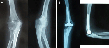
Figure 1
Plain radiographs of the right elbow of a 29-year-old female
patient (Case 1). (A) Preoperative anteroposterior and lateral views. (B)
Anteroposterior and lateral views after total elbow arthroplasty (TEA).
Figure 2
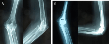
Figure 2
64-year-old female patient (Case 2). (A) Preoperative
anteroposterior and lateral views. (B) Anteroposterior and lateral views after
TEA.
Figure 3
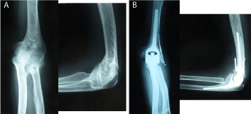
Figure 3
72-year- old female patient (Case 3). (A) Preoperative
anteroposterior and lateral views. (B) Anteroposterior and lateral views after
TEA.
Discussion
There have been several reported case series of TEA for ankylosed
elbows [3,7-9]. Figgies et al. [3] reported a 19-case series of TEA
using linked implants for ankylotic elbows in patients with a variety
of conditions that have included RA. Of the 19 cases, 4 cases were
evaluated as excellent, 11 as good, 3 as fair, and 1 as a failure. The
failed case occurred after an infection developed around the surgical
site and the implants were removed. The authors stated that the
functional limitations of patients should be evaluated at the outset
and this should include a concurrent evaluation of other upper
extremity joints. Mansat et al. [8] reported 14 cases, 4 of which were
rated as excellent, 4 were good, 1 was fair, and 5 were poor. Peden et
al. [9] reported 13 cases: 5 were rated as excellent, 3 were good, 4 were
fair, and 1 was poor. According to these reports, functional ability
restored in most patients postoperatively and was sufficient enough
that they could perform ADL normally. In our cases, two of the three
patients were very satisfied with the surgical outcome and reported
that their ADLs were better than they had been preoperatively.
In the previous reports, the range of motion achieved at the
elbows was mostly good. Figgie at al. [3] reported that the mean arc
of motion improved from 0 degrees preoperatively to 80 degrees
(range, 35 to 115 degrees of flexion) at an average 5.75 years after
the operation. Mansat et al. [8] reported that the mean increase in
the arc of flexion was 60 degrees (range, 5 to 115 degrees), with a
mean increase of 33 degrees in flexion and 27 degrees in extension.
Peden et al. [9] reported that the improvement in range of motion
at the elbow measured 1 year after the operation was maintained
for all patients after a 12-year follow-up. In our study, the average
arc of motion was 85 degrees (range, 65 to 110 degrees) at the most
recent examination. This result is similar to that of the Peden et al. [9]
study. Improvement in the range of motion of the elbow improves
the ADL for patients with RA. Common complications following
TEA are aseptic loosening, infections, ulnar nerve problems, elbow
instability, dislocation, subluxation, intraoperative fracture, fracture
of the prosthesis, and ectopic bone formation [10]. When performing
TEA for an ankylotic elbow, special care should be taken to prevent
infection, ulnar nerve problems, instability, and an intraoperative
fracture [3,8-10]. Infection is not typically frequent, but severe
complications can arise if it does occur. In the previous study reported
by Figgie et al. [3] the only case to fail was due to a deep infection
and the patient was not a suitable candidate for implant arthroplasty
because of noncompliance. Mansat et al. [8] reported that a deep
infection developed in one elbow of two patients with posttraumatic
stiffness. One patient had two previous operations prior to the
arthroplasty, and the other patient had been operated on four times before. Both of these patients required revision surgery. In another
previous report, three patients had deep perioperative infections that
required an average of four additional operations [9]. Neurogenic
symptoms, especially those pertaining to ulnar nerve disorders, were a
relatively common complication after TEA. It was reported that some
patients with moderate neurogenic symptoms preoperatively had
complete relief after ulnar nerve decompression and transposition,
while other previously asymptomatic patients developed ulnar nerve
symptoms; however, no pain was attributed to the ulnar nerve [8].
Among our cases, one of the three patients had significant ulnar
nerve symptoms after undergoing TEA and required revision surgery
(neurolysis). Because kinking or compression of the ulnar nerve can
occur after TEA for ankylotic elbows [3], perioperative procedures
to manage the ulnar nerve should be employed. In previous reports,
linked implants have been recommended in order to maintain the
stability of elbows over the full course of the period during which
they are observed [8,9]. Our cases had received linked implants and
we did not encounter complaints regarding symptoms caused by
elbow instability. Intraoperative fracture is one of the most severe
complications to try and avoid during this type of operation. Peden
et al. [9] reported that two cases had a fracture of the lateral humeral
epicondyle which resolved uneventfully and the ulnar component
was malpositioned causing a perforation of the posterior cortex in
another case. Mansat et al. [8] reported that two patients sustained
a fracture associated with a loose component. One of our cases had
an intraoperative fracture and it was fixed with a Kirchner wire.
We noted that the original border between the humerus and ulna
was sometimes difficult to identify intraoperatively and this can
inadvertently contribute to a fracture when opening up an ankylotic
joint.
In conclusion, TEA for ankylotic elbows in patients with RA is
a good surgical option to restore adequate function as well as any
other spontaneous ankylosis. For the sake of a good outcome, it is
necessary to evaluate other joints in the extremity in order to properly
evaluate the mobility and function of the elbow and to prepare a
comprehensive plan of pre- and post-operative management that
takes into consideration the full motion of the upper extremity.
References
- Momohara S, Inoue E, Ikari K, Ochi K, Ishida O, Yano K, et al. Recent trends in orthopedic surgery aiming to improve quality of life for those with rheumatoid arthritis: data from a large observational cohort. J Rheumatol. 2014; 41: 862-866.
- Souther WA. Surgery for rheumatoid arthritis: upper limb surgery of the elbow. Ann Rheum Dis. 1990; 49: 871-872.
- Figgie MP, Inglis AE, Mow CS, Figgie HE. Total elbow arthroplasty for complete ankylosis of the elbow. J Bone Joint Surg Am. 1989; 71: 513-520.
- O’Neill OR, Morrey BF, Tanaka S, An KN. Compensatory motion in the upper extremity after elbow arthrodesis. Clin Orthop Relat Res. 1992; 281: 89-96.
- Glynn JJ, Niebauer JJ. Flexion and extension contracture of the elbow: surgical management. Clin Orthop Relat Res. 1976; 117: 289-291.
- Weizenbluth M, Eichenblat M, Lipskeir E, Kessler I. Arthrolysis of the elbow. 13 cases of posttraumatic stiffness. Acta Orthop Scandinavica. 1990; 60: 642-645.
- Kraay MJ, Figgie MP, Inglis AE, Wolfe SW, Ranawat CS. Primary semiconstrained total elbow arthroplasty. Survival analysis of 113 consecutive cases. J Bone Joint Surg Br. 1994; 76: 636-640.
- Mansat P, Morrey BF. Semiconstrained total elbow arthroplasty for ankylosed and stiff elbows. J Bone Joint Surg Am. 2000; 82: 1260-1268.
- Peden JP, Morrey BF. Total elbow replacement for the management of the ankylosed or fused elbow. J Bone Joint Surg Br. 2008; 90: 1198-1204.
- Voloshin I, Schippert DW, Kakar S, Kaye EK, Morrey BF. Complications of total elbow replacement: a systematic review. J Shoulder Elbow Surg. 2011; 20: 158-168.