Case Report
Spontaneous Resolutıon of Stauffer’s Syndrome in Advanced Renal Cell Carcınoma: A Case Report
Ozlem Yersal1*, Nezih Meydan2, Sabri Barutca2
1Samsun Research and Training Hospital, Turkey
2Department of Oncology, Adnan Menderes University, Turkey
*Corresponding author: Ozlem Yersal, Samsun Research and Training Hospital, Baruthane Mahallesi, İskenderun Sokak, No:30 D:2, 55000, İlkadım/SAMSUN, Turkey
Published: 09 Dec, 2016
Cite this article as: Yersal O, Meydan N, Barutca S.
Spontaneous Resolutıon of Stauffer’s
Syndrome in Advanced Renal Cell
Carcınoma: A Case Report. Ann Clin
Case Rep. 2016; 1: 1211.
Abstract
Stauffer syndrome is characterized by abnormal liver function tests, hepato-splenomegaly, histologic changes consistent with non-specific hepatitis, and recovery of these abnormalities after nephrectomy, in patients with RCC. Few cases have been described with paraneoplastic cholestatic jaundance of RCC in the literature. We report a case of a female patient presenting with advanced renal cell carcinoma with Stauffer’s syndrome who has recovered spontaneously.
Keywords: Stauffer's syndrome; Renal carcinoma
Introduction
Renal Cell Carcinoma (RCC) is associated with an up to 10% to 40% prevalence of paraneoplastic
syndromes either at the time of diagnosis or during the disease course. Hypercalcemia, hypertension,
polycythemia, nonmetastatic hepatic dysfunction, galactorrhoea, Cushing's syndrome, alterations
in glucose metabolism, amyloidosis, anemia, neuromyopathy, vasculopathy, nephropathy,
coagulopathy and prostaglandin elevation are paraneoplastic manifestations, that has been described
among RCC patients [1].
In 1961, Stauffer described abnormal liver function tests, hepato-splenomegaly, histologic
changes consistent with non-specific hepatitis, and recovery of these abnormalities after
nephrectomy, in patients with RCC [2]. Cytokin production by the tumor, especially interleukin
6 are considered as the underlying pathogenetic mechanism of RCC patients who present with
hepatic dysfunction in the absence of liver metastases.
We report a case of an female patient presenting with advanced renal cell carcinoma with
Stauffer’s syndrome who has recovered spontaneously.
Case Presentation
A 69 year old female patient was admitted with jaundance and pruritis of two weeks duration.
She had a history of right radical nephrectomy nineteen years before and the final pathologic
examination had demonstrated clear cell carcinoma with renal vein invasion. Follow up magnetic
resonance imaging confirmed a left renal mass of 7 cm, thirteen years after operation. The patient
refused surgery and any medical treatment and was on radiologic follow up. However, after a follow-up period of two years, renal mass increased in size and she
developed a second renal 5 cm mass. She received interferon-alpha
therapy until radiologic evidence of disease progression for three
months. Sorafenib was administered as second line treatment which
was discontinued by the patient because of severe skin reactions,
while the disease regressed. She again denied treatment and lost from
follow up.
Physical examination revealed jaundence, hepatomegaly and a
large palpable, relatively mobile, nontender mass in the left side of
the abdomen. Laboratory data revealed anemia leucocytosis, elevated
alkaline phospatase (ALP), gamma-glutamyl transpeptidase (GGT),
total billurubin, calcium, decreased serum albumin levels and
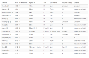
prolonged prothrombine time (Table 1). Protein electrophoresis
revealed mild diffuse hyperglobulinaemia. She was negative for
hepatitis A,B,C,D, cytomegalovirus, Epstein-Barr virus, herpes virus
serological tests. Immunological assays for the various autoimmune
diseases (ANA, ENA, antids DNA, antiss DNA, antihistones
antibodies, ASMA, AMA, p-ANCA, c-ANCA) were all negative.
The urine analysis showed hematuria, blood and urine cultures were
sterile.
Abdominal ultrasonography showed 94 and 75 mm masses on
the left kidney. There was no evidence of intrahepatic or extrahepatic
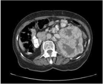
biliary dilatation. Abdomen computed tomography showed two
masses on left kidney and multipl paraaortic lymphadenopathies
(Figure 1). MRCP revealed normal pancreas and no intra or
extrahepatic biliary dilatation. The patient's liver function test levels
gradually increased reaching the highest values one month from
admission (Table 1).
The patient denied liver biopsy and only treated with
cholestyramine supportively. On follow up billurubin, ALP and GGT
levels gradually decreased and became in normal ranges 3 months
later without any therapy (Table 1). She is on follow up without any
treatment for one year and her billurubin level is in normal ranges.
Figure 1
Discussion
Malignancy related cholestasis is usually associated with
mechanichal obstruction of biliary tract or widespread hepatic
metastasis. It can rarely be the paraneoplastic manifestation of some
malignancies; especially renal cell carcinoma [3].
Few cases have been described with paraneoplastic cholestatic
jaundance of RCC in the literature [4]. Our patient had elevated ALP, GGT and billurubin levels, decreased albumin and prolonged
prothrombin time. There was no liver abnormality on abdomen
computed tomography. On the basis of these findings and absence
of obvious cause of liver dysfunction the patient diagnosed as having
Stauffer’s syndrome.
Stauffer’s syndrome with cholestatic jaundence, appears as the
initial clinical presentation of renal cell carcinoma. All described
patients were diagnosed during an evulation of jaundence [5]. They
all underwent nephrectomy and had complete resolution of clinical
and liver function abnormalities after surgery. Recently, Girotra et al
reported a patient with prior nephrectomy who developed Stauffer’s
syndrome as a presentation of RCC recurrence [6]. The patient denied
further workup and there is no information about clinical course
thereafter. Authors concluded that this syndrome may be a surrogate
marker of RCC recurrence. Our patient had prior nephrectomy for
RCC and she had developed a mass on the other kidney on follow
up. Stauffer’s syndrome diagnosis was not obtained neither prior the
diagnosis of RCC nor before recurrence. Jaundence occurred after
disease recurrence when she was not taking any medications for
RCC. She was icteric on admission, billurubin levels tend to decrease
and became normal levels 3 months after. Resolution of Stauffer’s
syndrome was spontaneously as she did not accept operation and any
other medication for RCC.
The underlying pathophysiology of
this paraneoplastic manifestation is poorly understood. However
possible role of IL-6 overexpression by the primary tumor are
reported. Blay et al. [7] investigated this issue in 119 metastatic renal
cell carcinoma patients and found that patients with detectable IL-6
had significantly higher serum ALP and GGT levels. They concluded
that IL-6 is involved in the pathophysiology of paraneoplastic
cholestasis. Think that our case adds to the growing pool of data of
this rare variant of Stauffer’s syndrome. There is no data about non
treated paraneoplastic cholestatic jaundence of RCC in the literature.
This is the first case of spontaneous resolution of this paraneoplastic
syndrome.
Table 1
References
- Palapattu SG, Kristo B, Rajfer J. Paraneoplastic Syndromes in Urologic Malignancy: The Many Faces of Renal Cell Carcinoma. Rev Urol. 2002; 4: 163-170.
- Stauffer MH. Nephrogenic hepatosplenomegaly. Gastroenterology. 196; 40: 694.
- Gold P, Fefer A, Thompson J. Paraneoplastic manifestations of renal cell carcinoma. Semin Urol Oncol. 1996; 14: 216-222.
- Dourakis SP, Sinani C, Deutsch M. Cholestatic jaundice as a paraneoplastic manifestation of renal cell carcinoma. Eur J Gastroenterol Hepatol. 1997; 9: 311-314.
- Giannakos G, Papanicolaou X, Trafalis D. Stauffer’s syndrome variant associated with renal cell carcinoma. Int J Urol. 2005; 12: 757-759.
- Girotra M, Abraham RR, Pahwa M, Arora M. ANZ Journal of Surgery. 2010; 80: 949-950.
- Blay JY, Rossi JF, Wijdenes J. Role of IL-6 in paraneoplastic inflammatory syndrome associated with renal cell carcinoma. Int J Cancer. 1997; 72: 424- 30.


