Case Report
Case of a Large Unruptured Sinus of Valsalva Aneurysm Invoving Non Coronary Cusp
Malini Sasidharan, Ajay Alex* and Harikumaran Nair
Department of Radio Diagnosis, Government T.D. Medical College, India
*Corresponding author: Ajay Alex, Department of Radio diagnosis, Government T.D. Medical College, Alappuzha, Kerala, India
Published: 09 Dec, 2016
Cite this article as: Sasidharan M, Alex A, Nair H. Case of
a Large Unruptured Sinus of Valsalva
Aneurysm Invoving Non Coronary
Cusp. Ann Clin Case Rep. 2016; 1:
1209.
Abstract
A sinus of Valsalva aneurysm is a rare cardiac anomaly that may be congenital or acquired. It involves aneurysmal dilation of the coronary or non coronary cusps at the region of aortic root. These are more common to involve the coronary cusps, with very few case reports of an unruptured non coronary cusp involvement. We report a case of a 58 yr old female presenting with dyspnoea and chest pain. Clinical examination revealed bilateral pedal oedema and pan systolic cardiac murmur. Chest radiograph and echocardiography was done for the patient with contrast enhanced computed tomography (CECT) and Magnetic Resonance Imaging (MRI) done for detailed evaluation. Imaging showed a large unruptured sinus of Valsalva aneurysm involving the non coronary cusp of aorta compressing the left atria and RVOT. Thrombosis of the wall of the aneurysm was also identified. Large sinus of Valsalva aneurysms which remain unruptured, are rare as per literature. Patient underwent patch repair of the lesion and was put on follow up.
Keywords: Sinus of valsalva aneurysm; Cardiac MRI; Cardiac failure
Introduction
A sinus of Valsalva aneurysm is a rare cardiac anomaly that may be congenital or acquired; a coexisting cardiac lesion might be present. Non ruptured aneurysms may be asymptomatic and incidentally discovered, or they manifest acutely with mass effect on adjacent cardiac structures. Ruptured Valsalva sinus aneurysms result in an aorto-cardiac shunt and manifest as acute chest pain, dyspnoea and progressive congestive heart failure, or cardiac arrest. These are more common to involve the coronary cusps, with very few case reports of an unruptured non coronary cusp involvement. Prompt and accurate diagnosis is critical as both ruptured and non ruptured Valsalva sinus aneurysms may have potentially fatal complications. The treatment of choice is surgery. Most valsalva sinus aneurysms are diagnosed on the basis of echocardiography, with or without angiography. However, both contrast enhanced computed tomography and Magnetic Resonance (MR) imaging can provide excellent anatomic depiction. MR imaging can provide additional functional information. We present a case of patient with acute onset of symptoms and diagnosed as unruptured large sinus of Valsalva aneurysm.
Case Presentation
A 58-year-old woman presented with 1 month history of breathlessness, chest pain and
palpitation. On clinical examination patient had bilateral pedal oedema. Cardiac auscultation
revealed a pan systolic murmur in the aortic region. A chest radiograph was obtained (Figure 1). The
frontal radiograph obtained with the patient erect showed cardiomegaly with increased convexity
of right heart border. On transthoracic echocardiography, she was found to have a large blood filled
structure compressing left atrium posteriorly. Contrast enhanced CT study of thorax was done and
cardiac Magnetic Resonance (MR) imaging was performed few days later.
Contrast enhanced CT study of thorax showed a saccular out pouching of size 7.2 x 7.1 x 5.8 cm
arising from the aortic root on its postero-lateral side towards right (Figure 2). The aneurysmal wall
towards its medial aspect shows thrombosis of maximum thickness 1.6 cm and multiple areas of
discontinuous peripheral calcification. No definite communication of the sac with any of the heart
chambers could be demonstrated.
MRI showed a well defined saccular aneurysm arising from the non coronary sinus at the root
of ascending aorta on right side, with circumferential partial thrombosis (Figure 3). Aneurysm
was seen bulging into and moderately compressing the left atrium. Interatrial septum appears
displaced. Mild indentation noted on right atrium and right ventricle. SVC is laterally displaced and compressed by the aneurysm and is draining into the right atrium.
No SVC thrombosis. Compression of right pulmonary artery noted
which was seen draping over the superior aspect of the aneurysm.
Pulmonary trunk and left pulmonary artery is of normal calibre. The
right and left coronary cusps appear normal in morphology. The right
and left coronary artery appeared normal in course and calibre. Rest
of the aorta, arch and its branches are normal in enhancement and
morphology.
The patient underwent repair of the aneurysm with a prosthetic
patch. Patient was put on follow up post procedure.
Figure 1
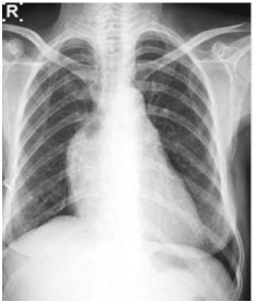
Figure 1
Chest radiograph PA view of 58-year old woman presented with
acute breathlessness showing mild cardiomegaly with increased convexity
of right heart border.
Figure 2
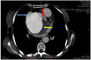
Figure 2
Contrast enhanced CT of thorax- axial section show out pouching
arising from the aortic root(red arrow) involving the non coronary cusp.SVC
(blue arrow) seen compressed laterally by the aneurysm. Lesion is seen
posteriorly compressing the left atria. Thrombus (yellow arrow) is noted
involving the wall of the aneurysm.
Figure 3
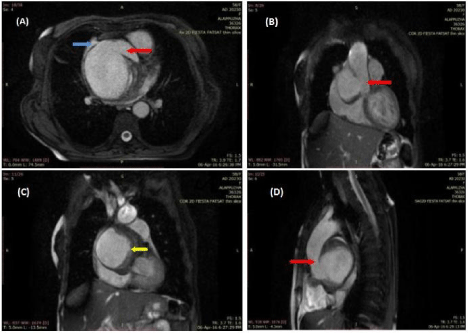
Figure 3
Cardiac MRI FIESTA sequence: (A) Axial section show out
pouching arising from the aortic root (red arrow) involving the non coronary
cusp.SVC (blue arrow) seen compressed laterally by the aneurysm; (B,C)
Coronal image showing the sinus of Valsalva aneurysm arising from the
aortic root; (D) Sagittal section showing the sinus of Valsalva aneurysm seen
arising posteriorly compressing left atria. Thrombosis (yellow arrow) involving
the aneurysm wall can also be seen.
Discussion
The sinus of Valsalva aneurysm is rare and accounts for about
0.15-3.5% of congenital heart diseases. These aneurysms are three to
four times as common in men as in women, and five times as common
in Eastern and Asian countries as in Western countries [1,2]. The
Valsalva sinuses are three subtle dilatations of the aortic root wall that
arise between the aortic valve annulus and the sinotubular ridge. Each
sinus is associated with a corresponding right, left or non coronary
aortic valve cusp. Valsalva sinus aneurysms can be congenital or
acquired. Congenital aneurysms occur due to fundamental localized
weakness of the elastic lamina at the junction of the aortic media
and the annulus fibrosis or associated with an underlying deficiency of normal elastic tissue, such as in Marfan and Ehlers-Danlos
syndromes. Acquired aneurysms commonly are caused by infectious
diseases such as bacterial endocarditis, syphilis and tuberculosis;
degenerative conditions such as atherosclerosis and cystic medial
necrosis; and injury from deceleration trauma.
The right sinus of Valsalva is most commonly involved (75% –
90%) [3]. The common cardiac anomalies that occur with Valsalva
sinus aneurysms are ventricular septal defect (supracristal type),
aortic insufficiency, bicuspid aortic valve and less frequently,
coronary anomalies. The characteristic diagnostic features of sinus
of Valsalva aneurysm include aneurysmal sac originating above the
aortic annulus, saccular shape of the aneurysm, and normal diameter
of the aortic root and ascending aorta [4].
When Valsalva sinus aneurysms rupture, they most commonly
rupture into the right ventricle, followed by the right atrium and
rarely into the left-sided heart chambers, pericardium and pulmonary
artery or superior caval vein [5].
Recently echocardiography has replaced angiography as the
principal technique to diagnose the aneurysm. In patients who have
suboptimal evaluation at echocardiography, CT and MR imaging
are useful to delineate sinus of Valsalva aneurysm. Both show the
structural appearance of the aneurysm, but MR imaging offers direct
multiplanar depiction without the need for injection of contrast
material. MR imaging shows turbulence within the aneurysm and
gradient-echo images may document aortic regurgitation. Cine
phase-contrast MR imaging delineates the site of fistula formation
and can be used to determine the extent of shunting caused by
rupture of the aneurysm [6]. The clinical manifestation of sinus of
Valsalva aneurysm varies widely. Unruptured aneurysms are mostly
asymptomatic and when symptoms are present are mostly related to
aneurysm rupture or mass effect on adjacent cardiac structures. Most
common presenting symptom is dyspnoea, seen in about 56% cases
and most common clinical sign is cardiac murmur.
Therefore, large unruptured aneurysms of the non-coronary
sinus of Valsalva may compress the adjacent mediastinal structures and patients may present with symptoms, similar to the present case. Very few cases of large unruptured sinus of Valsalva aneurysm
have been found in literature. The reported cases of sinus of Valsalva
aneurysm have usually been associated with ruptured aneurysm and
resultant fistula formation with right cardiac chambers or RVOT.
The mainstay of treating a case of symptomatic unruptured and
ruptured SVA is cardio-pulmonary bypass surgery with excision of
aneurysmal sac and the resultant defect will be repaired either by
direct suturing or patch closure. Coexisting lesions will be repaired
within the same surgical procedure [7]. Percutaneous transcatheter
closure can also be used for treating sinus of Valsalva aneurysm. In
asymptomatic patients surgery can be deferred if the aneurysmal sac
is small, however, large aneurysms should undergo surgical repair to
avoid complications.
Conclusion
Sinus of Valsalva aneurysm can involve coronary or non coronary sinus of aortic root. Large unruptured aneurysms are relatively rare but can present with cardiac failure and RVOT obstruction. Computed tomography and cardiac MR are essential investigations to depict the exact site of origin and extent of lesion in addition to rule out rupture. Surgery is the treatment of choice with patients having relatively good prognosis following repair.
References
- Mayer ED, Ruffmann K, Saggau W. Ruptured aneurysms of the sinus of Valsalva. Ann Thorac Surg. 1986; 42: 81-85.
- TakachTJ, Reul GJ, Duncan JM. Sinus of Valsalva aneurysm or fistula: management and outcome. Ann Thorac Surg. 1999; 68: 1573-1577.
- Smith WA. Aneurysm of the sinus of Valsalva with report of two cases. JAMA. 1914; 62: 1878.
- Ring WS. Congenital heart surgery nomenclature and database project: aortic aneurysm, sinus of Valsalva aneurysm, and aortic dissection. Ann Thorac Surg. 2000; 69: S147-S163.
- Feldman DN, Roman MJ. Aneurysms of the sinus of Valsalva. Cardiology. 2006; 106: 73-81.
- Edwards JE, Burchell HB. Specimen exhibiting the essential lesion in aneurysm of the aortic sinus. Proc Staff Meet Mayo Clin. 1956; 31: 407-412.
- Wang ZJ, Zou CW, Li DC, Li HX, Wang AB, Yuan GD. Surgical repair of sinus of Valsalva aneurysm in Asian patients. Ann Thorac Surg. 2007; 84: 156-60.