Clinical Image
Rhino-Orbito-Cerebral Mucormycosis with Three Intracranial Complications: Infarct, Hemorrhage and Ophthalmoplegia
Ali Jendoubi*
Department of Anaesthesia and Intensive Care, University Tunis El Manar, Tunisia
*Corresponding author: Ali Jendoubi, Department of Anaesthesia and Intensive Care, Charles Nicolle Hospital of Tunis, University Tunis El Manar, Tunis, Tunisia
Published: 08 Dec, 2016
Cite this article as: Jendoubi A. Rhino-Orbito-Cerebral
Mucormycosis with Three Intracranial
Complications: Infarct, Hemorrhage and
Ophthalmoplegia. Ann Clin Case Rep.
2016; 1: 1206.
Clinical Image
Rhino-orbito-cerebral mucormycosis is a rare life-threatening opportunistic fungal
infection caused by fungi from the order Mucorales [1]. The disease commonly affects
immunocompromised patients, especially ketoacidotic diabetic patients [2]. We report a case of
Rhinocerebral mucormycosis in a 48-year-old diabetic patient, who presented to our department
with the complaints of exophthalmos in the left eye, headache, fever and black nasal discharge
(Figure 1A and B). Magnetic Resonance Imaging (MRI) of the brain revealed left ethmoid sinusitis, leftside orbital cellulitis and suggested cavernous sinus thrombosis with left ophthalmic artery occlusion
(Figure 2 and Figure 3A). He developed progressive skin necrosis on the left periorbital region within 72 hours of admission (Figure 1C). Diagnosis of mucormycosis was confirmed by mycological findings. Initial empirical antimicrobial therapy was initiated with intravenous cefotaxime 2 g
intravenously (IV) every 8 hours, metronidazole 500 mg IV every 8 hours, fosfomycin 4 g IV every
6 hours and injectable amphotericin B. Continuous IV insulin infusion was administered with IV
fluids.
On the 4th day, the patient developed altered consciousness, the brain CT scan revealed a
cerebral hemorrhage in the left frontal lobe (Figure 3B). Despite aggressive therapy, patient succumbed to the disease.
This case illustrates the association of three lifethreatening
intracranial complications of mucormycosis: septic
cavernous sinus thrombosis with cerebral infarct, intracerebral
hemorrhage and ophthalmoplegia.
Intracranial dissemination of mucormycosis is associated with
increased mortality [2].
Infection spreads along vascular and neuronal structures
and infiltrates the walls of blood vessels. It spreads to the
contiguous sinuses and subsequently to the orbit, the retro-orbital
area and brain leading to brain abscess.
Involvement of the superior orbital fissure and its contents, such
as cranial nerves III, IV, and VI, and branches of V1 and V2, may
cause diplopia, ophthalmoplegia and blindness may occur [3].
A cerebral infarct may occur as a result of a thrombus or a mycotic
embolus after arterial invasion [4].
In this context, mucormycosis diagnosis is confirmed by the
presence of multiple irregular non-septate hyphae on histopathologic
examination [2].
Intracranial and intraorbital extension of mucormycosis is more
precisely demonstrated by MRI than by CT [5].
The basis of mucormycosis treatment remains a combination of
extensive surgical debridement and amphotericin B for a period of
4–6 weeks [6].
Prognosis may improve with rapid diagnosis, early management
and reversible underlying risk factors.
Figure 1
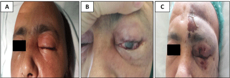
Figure 1
(A) ICU admission: Left palpebral oedema, local erythema, blindness and ptosis of the left eye. (B)
Left orbital cellulitis. (C) 72 hours after the admission: progressive skin necrosis on the left periorbital region.
Figure 2
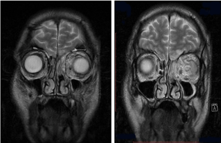
Figure 2
MRI brain: Left orbital cellulitis with contiguous spread of infection to the adjacent basifrontal region.
Figure 1
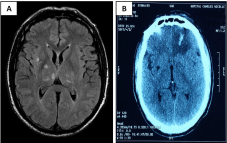
Figure 3
(A) MRI brain: ischemic lesions in the right caudate and lenticular
nuclei. (B) Head computed tomography scan showing an acute left frontal
hemorrhage.
References
- Spellberg B, Edwards J, Ibrahim A. Novel perspectives on mucormycosis: pathophysiology, presentation, and management. Clin Microbiol Rev. 2005; 18: 556-569.
- Bhansali A, Bhadada S, Sharma A, Suresh V, Gupta A, Singh P, et al. Presentation and outcome of rhino-orbital-cerebral mucormycosis in patients with diabetes. Postgrad Med J. 2004; 80: 670-674.
- Koc Z, Koc F, Yerdelen D, Ozdogu H. Rhino-orbital-cerebral mucormycosis with different cerebral involvements: Infarct, hemorrhage, and ophthalmoplegia. Int J Neurosci. 2007; 117:1677-1690.
- Chan LL, Singh S, Jones D, Diaz E M Jr, Ginsberg L E. Imaging of mucormycosis skull base osteomyelitis. American Journal of Neuroradiology. 2000; 21: 828-831.
- Ruoppi P, Dietz A, Nikanne E, Seppa J, Markkanen H, Nuutinen, J. Paranasal sinus mucormycosis: A report of two cases. Acta Otolaryngologica. 2001: 121: 948-952.
- EJC Goldstein, B Spellberg, TJ Walsh, DP Kontoyiannis, J Edwards Jr, A Ibrahim. Recent advances in the management of mucormycosis: from bench to bedside. Clin Infect Dis. 2009; 48: 1743-1751.