Case Report
Goltz-Gorlin Syndrome: Report of Two Cases
Rashid Soufizadeh* and Mozhgan Kazemian
Department of Oral and Maxillofacial Surgery, Mashhad University of Medical Sciences, Iran
*Corresponding author: Rashid Soufizadeh, Department of Oral & Maxillofacial Surgery, School of Dentistry, Mashhad University of Medical Sciences, Mashhad, Iran
Published: 07 Dec, 2016
Cite this article as: Soufizadeh R, Kazemian M. Goltz-
Gorlin Syndrome: Report of Two Cases.
Ann Clin Case Rep. 2016; 1: 1205.
Abstract
Goltz-Gorlin syndrome or Nevoid Basal Cell Carcinoma syndrome (NBCCS) is an infrequent
multisystemic disease that is inherited in a dominant autosomal pattern, which shows a high level
of penetrance and variable expressiveness. The syndrome is associated with multiple Keratocystic
Odontogenic Tumors (KCOT) in 90% of cases. In addition to jaw cysts, multiple basal cell
carcinomas and skeletal anomalies are common findings. KCOTs associated with this syndrome
can be seen in both maxilla and mandible but there is more tendencies to posterior mandible and
the recurrence rate is more than nonsyndromic cases.
In this article we describe two patients with diagnosis of Goltz-Gorlin syndrome, its typical findings
and finally we discuss about the treatment method used for jaw cysts and the outcome of 1 year
follow-up.
Keywords: Goltz-Gorlin syndrome; Nevoid basal cell carcinoma; Keratocystic odontogenic tumors
Introduction
Goltz-Gorlin syndrome or Nevoid Basal Cell Carcinoma syndrome (NBCCS) is an infrequent
multisystemic disease that is inherited in a dominant autosomal pattern, which shows a high level
of penetrance and variable expressiveness. In 1894, Jarisch and White made the first descriptions
of patients with this syndrome, highlighting the presence of multiple basal cell carcinomas. Then
in 1960, Robert W Goltz and Robert J Gorlin described a triad of signs of this disease including;
multiple nevoid basal cell carcinomas, multiple jaw keratocysts and bifid ribs [1,2]. Pathogenesis of
the syndrome is attributed to abnormalities in the long arm of chromosome 9 (q22.3-q31) and loss
of, or mutations of human patched gene (PTCH1 gene) [2].
The syndrome is associated with multiple Keratocystic Odontogenic Tumors (KCOT) in 90%
of cases. In addition to jaw cysts, multiple basal cell carcinomas and skeletal anomalies are common
findings. Skeletal findings are; bifid ribs, vertebral anomalies, bossing of frontal and temporoparietal
region, hypertelorism, palmar and plantar pit and fissures, Calcification of falx cerebri, ocular and
centeral nervous system lesions, cleft lip and palate, mandibular prognathism and in some rare cases
the ovarian fibroma [3]. KCOTs associated with this syndrome can be seen in both maxilla and
mandible but there is more tendencies to posterior mandible [4] and the recurrence rate is more
than nonsyndromic cases [5]. In this article we describe two patients with diagnosis of Goltz-Gorlin
syndrome, typical findings of it and finally we discuss about the treatment method used for jaw cysts
and the outcome of 1 year follow-up.
Case Presentation
Case 1
A 15 years old boy by a chief complain of pain in dental region referred to dentist and an
Orthopantomogram (OPG) asked for him. In the OPG of patient the multiple jaw lesions has been
diagnosed and patient has been referred to Mashhad dental faculty. In clinical examination, the
patient had a slight frontal and temporal bossing. There were no hypertelorism but he had unibrow.
There was no pigmented lesion in any region of patient’s body (Figure 1).
In OPG (Figure 2), radiolucent lesions related to bilateral mandibular and right maxillary
impacted third molars were observed. In the left maxilla second and third molars were impacted.
In the right mandible, radiolucent lesion with well defined and cortical border and large
extension from sigmoid notch to apical region of distal root of right 1st molar was observed that
led to displacement of impacted third molar to the gonial angle. Also in the left side of mandible
there was a well defined radiolucent lesion with cortical border in relation to impacted third molar that was 14*7mm dimensions. There was a slight tooth bud
displacement in this region. And in the right side of maxilla there
was a radiolucent, well defined with cortical border lesion related
impacted third molar that was caused the displacement of the tooth.
In the skull radiography, calcification of Falx cerebri was observed in
form of radiopaque lines in the center of the skull which is a typical
view of this syndrome (Figure 3).
Histopathological evaluation of jaw lesions revealed cystic wall
with ulcerated areas, squamous epithelium and wavy parakratine.
According to these findings, the diagnosis was Keratocystic
odontogenic tumor (KCOT).
Treatment plan for the patient was including the prophylactic
removal of impacted left maxillary teeth, removal of impacted third
molars with enucleation of related KCOT in the left mandibular and
right maxillary region and marsupialization of the right mandibular
cyst. Marsupialization was done by creating a window in the epithelium of cyst and suturing it to the surrounding periosteum and
then to keep it open by an acrylic obturator with a process extended
to the cyst was made. Patient followed every month and at third
month, an OPG was taken that revealed calcification of surgical site in
left side of mandible and decompression of cyst followed by upward
movement of impacted third molar in right side of mandible (Figure
4). No new lesions seen on the provided image.
Case 2
A 21 years old man with chief complains of pain and swelling
of lower jaw came and an OPG was ordered. In his OPG there were
multiple radiolucent lesions in both mandible and maxilla (Figure 5).
In clinical examination, the patient had a slight frontal bossing but
there was no hypertelorism. Also he had pigmented lesions in multiple
sites of his body. He had unibrow too (Figure 6). In the panoramic and
cone-beam computed tomography (Figure 7), there were radiolucent
lesions in 3 sites of jaws; right ramus and retromolar of mandible, left
retromolar of the mandible and distal of right maxillary 2nd molar.
There were no impacted teeth and the patient had no third molars.
In the right side of mandible, the radiolucent lesion was in the distal
of 2nd molar and posterior extension to mandibular ramus. The lesion
had a shape like clover leaves with well defined and cortical border
and approximate dimensions of 30*10mm that was caused bone perforation in retromolar region. In the left side of mandible, there
was a radiolucent lesion in the distal of 2nd molar with well-defined
and cortical border and approximate dimension of 16*20mm. This
lesion was extended posterior to the anterior of ramus. In the right
maxillary, a radiolucent lesion in the distal of 2nd molar with welldefined
and cortical border and approximate dimension of 28*25mm.
This lesion was extended anteroposteriorly and in the anterior
extension, was invaded into the maxillary sinus. Also, buccal cortex
of maxilla in distal of 2nd molar was destroyed completely. PA skull
radiography was obtained in which the calcification of falx cerebri
was observed (Figure 8) and in the chest radiography, there was no
bifid rib (Figure 9).
Surgery of jaw lesions were performed under general anesthesia.
In maxilla the radiolucent lesion enucleated completely and due
to involvement of surrounding bone, the 2nd molar extracted too.
In mandible, the treatment plan was to marsupialization of cystic
lesion in both sides, so a window in the epithelium of cystic lesions
was made and kept open by using an acrylic obturator in both sides.
Histopathologic diagnosis of all three regions was KCOT.
Also we did a biopsy from skin lesion of facial region and the
histopathologic diagnosis of it was Basal Cell Carcinoma (BCC).
The patient followed up monthly and OPG taken at 3rd month after
surgery showed the calcification in right maxillary region and reduce
in size of mandibular lesions. The patient referred to a dermatologist
for treatment of skin lesions.
Figure 1
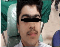
Figure 1
No pigmented lesion in any region of patient’s body.
Figure 2
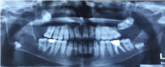
Figure 2
In OPG, radiolucent lesions related to bilateral mandibular and right
maxillary impacted third molars were observed. In the left maxilla second and
third molars were impacted.
Figure3
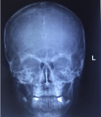
Figure 3
The skull radiography, calcification of Falx cerebri was observed
in form of radiopaque lines in the center of the skull which is a typical view
of this syndrome.
Figure 4
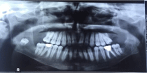
Figure 4
H & E, 10x, showing features of basal cell carcinoma with melanin
pigmentation at numerous areas.
Figure 5
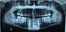
Figure 5
OPG there were multiple radiolucent lesions in both mandible and
maxilla.
Figure 6
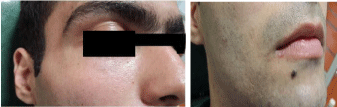
Figure 6
Panorama, 3-D, tangential and axial sections of Cone beam
computed tomography reveals absence of bony changes in the region of
interest and only radiopacity in the left maxillary sinus can be appreciated as
an incidental finding.
Figure 7
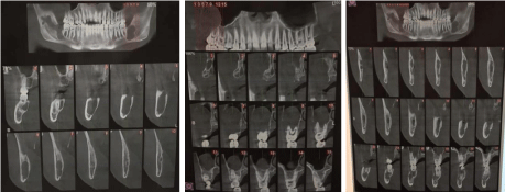
Figure 7
From left (Left mandibular cyst, right maxillary cyst and right mandibular cyst).
Figure 8
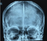
Figure 8
PA skull radiography was obtained in which the calcification of falx
cerebri was observed.
Figure 9
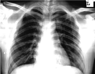
Figure 9
In the chest radiography, there was no bifid rib.
Discussion
The Gorlin syndrome has diagnostic features such as multiple jaw
KCOTs, NBCC, calcification of falx cerebri, palmar and plantar pit
and fissure, bifid ribs, hypertelorism, frontal and temporal bossing
and etc [6] there were KCOT in two side of mandible in our first case
and two side of mandible and right side of maxilla in the second case
and also there was calcification of falx cerebri in both cases. These
findings were similar to the diagnostic findings of Kavoosi and
colleagues [7]. According to Habibi and colleagues average age of
diagnosis of Gorlin syndrome in Iranian population is 35/12 years
[8]. which in our cases it was lower (15 years old in case 1 and 21 years
old in case 2). Multiple KCOTs is the primary diagnostic symptom of
this syndrome, as in our both cases.
According to Habibi and colleagues, there were multiple KCOTs
in all of 19 cases they studied [8] according to Gonzalez et al. [9]. These cysts are more willing to engage mandible. Which It was observed
in our patients too (Figure 1 and 6). According to Habibi et al [8], the calcification of falx cerebri exists in 89% of Iranian population
suffering from this syndrome which was observed in our both
patients. Multiple Basal cell carcinoma usually appears in puberty
or the second and third decades of life [9], in our first case there
was no skin lesion but the second patient had multiple skin lesions
on his face and upper limb that biopsy from facial lesions revealed
BCCs. KCOT treatment depends on several factors, including age,
size and location and extension of lesion, perforation of cortical bone and invasion to soft tissue [6,10]. According to this, treatments
are generally classified as conservative or aggressive; Conservative
treatment generally includes simple enucleation- with or without
curettage, or marsupialization. Aggressive treatment generally
includes peripheral ostectomy, chemical curettage with Carnoy’s
solution, cryotherapy, or electrocautery and resection [10]. Different
recurrence rates have been reported with each of these methods.
According to study of Kaczmaryc et al. [11] on the choice of treatment
method and recurrence rate of jaw lesions, the lowest recurrence rate
was in resection and the highest rate was in enucleation (with or
without adjunct therapy). Also, according to Titinchi and colleagues,
recurrence rate of KCOT is reported 29/2% that in their study, the
highest recurrence was associated with marsupialization [12]. In our
first patient that we did enucleation in right maxilla and left mandible
and marsupialization in right side of mandible for treatment of
KCOT, in 10 month follow-up period, there was no recurrence and
the size of right mandibular lesion was reduced.
In 18 months follow-up period of our second patient, there was
no recurrence in maxilla which was treated by enucleation. In the
mandibular lesions, according to OPG radiographies taken 3,6 and
12 months after surgery,the lesions was progressing towards healing
but in OPG taken at month 18, the radiolucency was a little increased.
So we requested a CBCT from both side of mandible and according to
it, lesion’s size was reduced but the cysts needed enucleation that was
done by a second surgery.
Conclusion
Gorlin-Goltz syndrome is a rare autosomal dominant disorder that involves multiple organ systems and if it be diagnosed as early as possible, there is the possibility of more effective treatment and prevention of some findings. So in suspected cases of this syndrome, Counseling and referral to other related specialties for a more comprehensive diagnostic evaluation is recommended.
References
- Agrawal A, Murari A, Vutukuri S, Singh A. Gorlin-Goltz Syndrome: Case Report of a Rare Hereditary Disorder. Case Rep Dent. 2012; 2012: 4.
- Joshi PS, Deshmukh V, Golgire S. Gorlin-Goltz syndrome. Dent Res J. 2012; 9: 100-106.
- Dixit S, Acharya S, Dixit PB. Multiple odontogenickeratocysts associated with Gorlin-Goltz syndrome. KUMJ. 2009; 7: 414-418.
- Melo ES, Kawamura JY, Alves CA, Nunes FD, Jorge WA, Cavalcanti MG. Imaging modality correlations of an odontogenickeratocyst in the nevoid basal cell carcinoma syndrome: a family case report. Oral Surg Oral Med Oral Pathol Oral Radiol Endod. 2004; 98: 232-236.
- Fonseca RJ, Marciani RD, Turvey TA. Oral and Maxillofacial Surgery: Trauma, surgical pathology, temporomandibular disorders: Saunders/ Elsevier; 2009.
- Kiwilsza M, Sporniak-Tutak K. Gorlin-Goltz syndrome--a medical condition requiring a multidisciplinary approach. Medical science monitor: International medical journal of experimental and clinical research. 2012; 18: Ra145- Ra153.
- Kavoosi A, Afghahi M, Hafezi L, shahsavari F, Sadri D. Basal cell nevus syndrome (Gorlin-Goltz) : A Case Report. J Res Den Sci. 2012; 9: 50-56.
- Habibi A, Jafarzadeh H. Nevoid basal cell carcinoma syndrome: a 17-year study of 19 cases in Iranian population (1991–2008). J Oral Pathol Med. 2010; 39: 677-680.
- Gonzalez-Alva P, Tanaka A, Oku Y, Yoshizawa D, Itoh S, Sakashita H, et al. Keratocystic odontogenic tumor: a retrospective study of 183 cases. J Oral Sci. 2008; 50: 205-212.
- Abdullah WA. Surgical treatment of keratocystic odontogenic tumour: A review article. Saudi Dent J. 2011; 23: 61-65.
- Kaczmarzyk T, Mojsa I, Stypulkowska J. A systematic review of the recurrence rate for keratocystic odontogenic tumour in relation to treatment modalities. Int J Oral Maxillofac Surg. 2012; 41: 756-767.