Case Report
Management of an Accessory Bile Duct Injury after Laparoscopic Cholecystectomy: A Case Report
Pierre Jean Aurelus*, Marco Salazar Domínguez and José Raúl Vázquez Langle
Hospital de Pediatría Centro Médico Nacional Siglo XXI, Mexico
*Corresponding author: Pierre Jean Aurelus, Hospital de Pediatría Centro Médico Nacional Siglo XXI (Instituto Mexicano del Seguro Social “IMSS”), Mexico
Published: 24 Nov, 2016
Cite this article as: Aurelus PJ, Domínguez MS, Langle
JRV. Management of an Accessory
Bile Duct Injury after Laparoscopic
Cholecystectomy: A Case Report. Ann
Clin Case Rep. 2016; 1: 1189.
Abstract
Background: Bile duct injury is a severe and potentially life–threatening complication of
laparoscopic cholecystectomy and the most difficult to resolve if there is an accessory bile duct.
This is a complex problem, where inadequate reconstruction has an impact on the quality of life of
patients. Some series have reported a 0.5% to 1.4% incidence of bile duct injury during laparoscopic
cholecystectomy. The aim of this case was to analyze the presentation, characteristics and treatment
results of an infant with an accessory bile duct injury after a laparoscopic cholecystectomy.
Case Presentation: A child of 13-year-old, male patient was referred to our center (Centro Medico
Nacional Siglo XXI: IMSS) for the management of cholelithiasis by laparoscopic cholecystectomy. In
his medical history, he had diffused abdominal pain while 2 years ago, ultrasound (US) that revealed
cholelithiasis (at least ten gallstone of different diameter 0.5 to 1cm), and an elective laparoscopic
cholecystectomy was performed. Ten days after, he presented a bile duct injury that we had been
repaired by PDS 6-0 and ferulization.
Conclusion: The cholelithiasis is not so frequently in infant and in child pathology, it is important
to evaluate hilar stricture to exclude the possibility of an accessory bile duct by a magnetic resonance
cholangiography (MRC) before the procedure. When we have involvement in the possibility of bile
duct injuries is better realized an abdominal scan and try to repair the bile duct by PDS 6-O by using
a catheter like ferulization in the first time before realized the Roux- en-Y choledocojejunostomy.
Keywords: Accessory bile duct-biliary tract injury; Laparoscopic cholecystectomy; Choledocojejunostomy
Introduction
Bile duct injury is a severe and potentially life–threatening complication of laparoscopic
cholecystectomy and most difficult to resolve if there is an accessory bile duct [1]. This is a
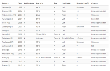
complex problem, where inadequate reconstruction has an impact on quality of life of patients [2]. Gurusamyl et al. [3] and others studies have reported a 0.5% to1.4% incidence of bile duct injury
during laparoscopic cholecystectomy and during the open cholecystectomies, the prevalence of
bile duct injury has been estimated at only 0.1-0.2 like difference (Table 1) [3-5]. Intrahepatic and
extrahepatic bile duct variations are commonly seen. The incidence of aberrant bile duct injury
associated with laparoscopic cholecystectomy has not yet been adequatelyrevised; abnormal biliary
anatomy is seen in large percent in the normal population [6-8]. It is important to visualize and make sure the site crossing of right hepatic artery by consideration of biliary duct [2]. Bile duct injury after laparoscopic cholecystectomy can be divided into the following categories: 1-the classic
injury; 2-variants of the classic injury; 3- burn injury; and 4-more remediable injuries [1].
Bile duct injury is a severe and potentially life –threatening complication of laparoscopic
cholecystectomy and most difficult to resolve if there is an accessory bile duct [1]. This is a complex problem, where inadequate reconstruction has an impact on quality of life of patients [2]. The management of patients following major bile duct injury is a surgical challenge often requiring
the skills of experienced hepatobiliary surgeons at tertiary referral centers [9]. In those injuries, the
most important, it is the repair procedure; like Sarmiento had evidenced that the life quality is the
same like a health patient after a good reconstruction of a hilar duct [2]. Major biliary injuries are more severe than traditional cholecystectomy and require multidisciplinary expertise for successful
results [1,2].
The aim of this case was to analyze the presentation, characteristics and treatment results of an
infant with an accessory bile duct injury after a laparoscopic cholecystectomy.
Table 1
Table 2
Case Presentation
A child of 13-year-old, male patient was referred to our center
(Centro Medico Nacional Siglo XXI: IMSS) for the management of
cholelithiasis by laparoscopic cholecystectomy. In his medical history,
he had diffuse abdominalpain while 2 years ago, without etiology and
no haematologic disease had been reported, only theultrasound (US)
revealed cholelithiasis (atleast tengall stones of different diameter 0.5
to 1 cm), and an elective laparoscopic cholecystectomy was performed
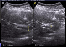
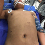
by using technical of three ports [10] (Figure 1 and 2).
The duration of laparoscopic cholecystectomy was 85 minutes
while the procedure was completed by three ports (generallywe
preferthe laparoscopic cholecystectomy by three ports, only, if, it
is necessary we use the fourth port), during the procedure we had
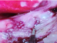
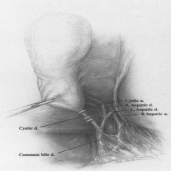
founded the right hepatic artery (RHA)across from behind the cystic
duct (CD) and there is no record of intraoperatively identified biliary
injury (Figure 3 and 4) and the patient was living home without
disturbance, ten days postoperative, he has had abdominal pain.
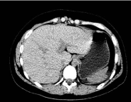
We realized an US without evidential abdominal collection. Six
hours after we performed an abdominalscan (Figure 5) and we have
identified a pelvic collection.
We performed a laparoscopic revision and we have identified
an accessory bile duct and we converted the procedure by an open
procedure, the remnant of cystic duct has identified and we have
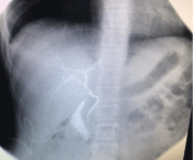
placed a catheter follow right hepatic duct and we have realized a
cholangiography, by identified the accessory duct we have closed it
by PDS 6-0, and the catheter had not removed for two months before
removing it, the patient has had no complains and the liver function
tests results within the normal limits (Figure 6).
Figure 1
Figure 2
Figure 3
Figure 4
Discussion
Since the introduction of laparoscopic cholecystectomy in 1987
by Philippe Mouret in France, an increase in these iatrogenic injuries
has been observed worldwide [4]. The laparoscopiccholecystectomy
is the preferred method for removing the gallbladder in the United
States. As with traditional open cholecystectomy, bile duct injury is
the most feared complication related to the new procedure [11]. The
biliary fistula is the most important injury in this procedure. There
have been a few proposals to classify postoperative strictures and
bile duct injuries. The Corlette-Bismuth classification is based on the length of the proximal biliary stump but not on the nature and length
of the lesion. Using this classification our patient had been injury type
five that is a combined common hepatic and aberrant right hepatic
duct injury, separating from the distal common bile duct (Table 2 and
Figure 3). In our patient if we had used the classification proposed by
McMahon, we had considered it like a major injury [1,2,4].
Biliary anatomical variations are encountered in 18.39% of
cases, with potentially hazardous anomalies predisposing to BTI in
only 3-6%. Anomalous right hepatic ducts are considered the most
dangerous type of anomaly, and our patient we had observed an
anomalous right hepatic duct (Figure 6) [4,12]. By the other hand, abnormal biliary anatomy, such as a short cystic duct or a cystic duct entering into the right hepatic duct would increase the incidence of
injuries in the bile duct [13].
Sometimes it is difficult to obtain the exact incidence rate
iatrogenic bile duct injury when there is an accessory duct not
identified previous the procedure, like our patient and when there
is not observed bile flow during the procedure. At the same time,
some Authors have also stressed the importance of an anomaly in
the right hepatic arterial running parallel to the cystic duct such as an
anomalous or accessory right hepatic artery. Common mechanisms of
injury during laparoscopic cholecystectomy are: 1- misidentification
of the cystic duct and the common hepatic duct because the cystic
duct is short (defined as cystic duct having a length of less than 5mm);
2-lateral clipping of the common hepatic duct; 3-traumatic avulsion
of the cystic duct junction, 4-diatermic injury of common hepatic
duct [9,8,12].
In this case the patient had presented some problems ten days
postoperative and the most important trouble had been an abdominal
diffuse pain. We had not imagined injury of an accessory bile duct,
because during the procedure we had been much emphasis to complete
the exposure of the peritoneal attachments in Carlot’s triangle and
the anatomical variations observed it had the right hepatic artery
coursing behind the cystic duct and we had not identified confluence
of any abnormal ducts into the cystic duct. However, during the
laparoscopic exploration, we had observed very clear the bile flow
by another duct. The management were included to open the cystic
duct and introduced a catheter to right hepatic duct and closed the
accessory bile duct by PDS 6-O in the right hepatic bile duct.
Some studies mentioned that during cholecystectomy, the
anatomical structure of Carlot’s triangle is not very clear because
of congestion, edema and fragility of the tissues around the cystic duct in acute suppurate or gangrenous cholecystic. Fibrous tissue
scars are often formed in Carlot’s triangle in atrophic cholecystitis
and it is more difficult to avoid intraoperative bile duct injuries, in
such conditions when correct identification of Carlot’s triangle is less
likely, intrahepatic bile duct anatomy is complex with many common
and uncommon variations. In spite of excellent laparoscopic
visualization complications. Perioperative lesions vascular structure
or extrahepatic (especially accessory) bile ducts during laparoscopic
cholecystectomy are a frequent cause of intra- and –postoperative
injury. The most common variant in the Radha Sarawagi study was
right posterior sectoral duct draining into the left hepatic duct in
27.6% of subjects [1,14-17].
In our patient it was different even-though it very important to
realize a magnetic resonance in pediatric patient with cholelithiasis
because it is not frequently like an adult this pathology in infant and
one of the diagnostic suspect had included hilar bile abnormality,
cholangiopancreatography (MRCP) is an excellent non-invasive
imaging technique for visualization of detailed biliary anatomy
[8,16]. It is our contribution in this case. The other importation in this
case is by the suspect of injury after laparoscopic cholecystectomy,it
is better realizing an abdominal scan.In this patient, like the accessory
duct was identifiedwith a grand possibility to close the bile flow it
had been not necessary a hepaticojejunostomyby Roux –en-Y jejunal
limb, or less commonly an end to side Roux-Y choledocojejunostomy
[1,11,8,15].
Figure 5
Figure 6
Conclusion
The cholelithiasis is not so frequentlyin infant and in child pathology, it is important to evaluate hilar stricture o exclude the possibility of an accessory bile duct by a magnetic resonance cholangiography (MRC) before the laparoscopic cholecystectomy procedure. When we have involvement, in the possibility of bile duct injuries, it is better realized an abdominal scan and try to repair the bile duct by PDS 6-O by using a catheter like ferulization in the first time, before realized the Roux- en-Y choledocojejunostomy. Expert surgeons have stressed the importance to open calot’s triangle, thereby reducing the likelihood of misidentification. Clear visualization of both: cystic duct and the choledochus, should be obtained before clip placement and transection of the cystic duct. Overuse of electrocautery must be avoided during the dissection of calot’s triangle because the heat transduction should be caused no identified injury during the procedure.
Acknowledgement
The Author would like to refer especial thanks to his family.
References
- G Branum, C Schmitt, J Baillie, P Suhocki, M Baker, A Davidoff, et al. Management of major biliary complications after laparoscopic cholecystectomy. Annsurg. 1993; 217: 532-541.
- Hector Losada M, Cesar Muñoz C, Luis Burgos S. Reconstrucción de lesión de la vía biliar principal. La evolución hacia la técnica de hepp-cuinaud. Rev Chil Cir. 2011; 63: 48-53.
- Gurusamy K, Samraj K, Gluud C, Wilson E, Davidson BR. Meta-analysis of randomized controlled trials on the safety and effectiveness of early versus delayed laparoscopic cholecystectomy for acute cholecystitis. Br J Surg. 2010; 97: 141-150.
- Viste A, Horn A, Øvrebø K, Christensen B, Angelsen JH, Hoem D. Bile duct injuries following laparoscopic cholecystectomy. Sage journal. 2016: 1-9.
- Balija M, Huis M, Szerda F, Bubnjar J, Stulhofer M. Laparoscopic cholecystectomy- accessory bile ducts. Acta Med Croatica. 2003; 57: 105- 109.
- Jirasiritham J, Wilasrusmee C, Poprom N, Larbcharoensub N. Pancreaticobiliary Ductal Anatomy in the Normal Population. Asian Pac J Cancer Prev. 1999; 17: 463-465.
- Uchiyama K, Tani M, Kawai M, Ueno M, Hama T, Yamaue H. Preoperative evaluation of the extrahepatic bile duct structure for laparoscopic cholecystectomy. G chir. 2013; 34: 249-253.
- Sureka Binit, Bansal Kalana, Patidar Yashwant, Arora Ankur. Magnetic resonance cholangiographic evaluation of intrahepatic and extrahepatic bile duct variations. Indian J Radiol Imaging. 2016; 26: 22-32.
- Parmeggiani D, Cimmino G, Cerbone D, Avenia N, Ruggero R, Gubitosi A, et al. Biliary tract injuries during laparoscopic cholecystectomy: three case reports and literature review. G Chir. 2110; 31: 16-19.
- Agrusa A, Romano G, Cucinella G, Cocorullo G, Bonventre S, Salamone G, et al. Laparoscopic,three-port and SILS cholecystectomy: a retrospective study. G Chir. 2013; 34: 249-253.
- National Institution of Health consensus development conference on gallstones and laparoscopic cholecystectomy. Bethesda, Maryland. 1992; 10: 1-20.
- Yamamoto S, Sakuma A, Rokkaku K, Nemoto T, Kubota K. Anomalous connection of the right hepatic duct into the cystic duct: utility of magnetic resonance cholangiopancreatography. Hepatogastroenterology. 2003; 50: 643-644.
- Wu YH, Liu ZS, Mrikhi R, Ai ZL, Sun Q, Bangoura G, et al. Anatomical variations of the cystic duct two case reports. World J Gastroenterol. 2008; 14: 155-157.
- Idu M, Jakimowicz J, Iuppa A, Cuschieri A. Hepatobiliary anatomy in patients with transposition of the gallbladder: implications for safe laparoscopic cholecystectomy. Br J Surg. 1996; 83: 1442-1443.
- Cuschieri A, Dubois F, Mouiel J, Mouret P, Becker H, Buess G, et al. The European experience with laparoscopic cholecystectomy. Am J Surg. 1991; 161: 385-387.
- Sarawagi Radha, Sundar Shyam, Raghuvanshi Sameer, Gupta Sanjeev Kumar, Jayaraman G. Common and uncommon variants of intrahepatic bile ducts in magnetic resonance cholangiopancreatography and its clinical implication. Pol J Radiol. 2016; 81: 250-255.
- Balija M, Huis M, Szerda F, Bubnjar J, Stulhofer M. Laparoscopic cholecystectomy-accessory bile ducts. Acta Med Croatica. 2003; 57: 105- 109.

