Case Report
Thyroid Paraganglioma: An Extremely Rare Tumor of the Thyroid
Adnan Özpek1*, Gözde Kir2, Hüseyin Kerem Tolan1, Metin Yucel1, Kemal Tekesin3, Gürhan Bas1 and Orhan Alimoglu4
1Department of General Surgery, Umraniye Training and Research Hospital, Turkey
2Department of Pathology, Umraniye Training and Research Hospital, Turkey
3Department of General Surgery, Usküdar State Hospital, Turkey
4Department of General Surgery, Istanbul Medeniyet University, Turkey
*Corresponding author: Adnan Özpek, Ümraniye Training and Research Hospital, Adem Yavuz Caddesi, No:1, 34766 Ümraniye, İstanbul, Turkey
Published: 24 Nov, 2016
Cite this article as: Özpek A, Kir G, Tolan HK, Yucel
M, Tekesin K, Bas G, et al. Thyroid
Paraganglioma: An Extremely Rare
Tumor of the Thyroid. Ann Clin Case
Rep. 2016; 1: 1188.
Abstract
Background: Paraganglioma originates from the para-ganglionic system which is mostly located in the adrenal gland medulla is a rare condition seen in the thyroid gland accounting for the 0.1% of
all the thyroid malignancies.
Case Presentation: A 67 years old female patient was admitted to our out-patient clinic with a
swelling around the neck and an unexplained chronic cough. The ultrasound (US) revealed a 33 x
20 mm hypoechoic nodule on the right lobe of the thyroid gland without any cervical lymph node
enlargement. Fine Needle Aspiration Biopsy (FNAB) was performed from the dominant nodule with
the guidance of US. In the cytopathology report there was a suspicion of neuro-endocrine tumor. In
the operation the nodule was found to be fixed to the adjacent trachea and a Total Thyroidectomy
was performed to the patient. Finally, in the histopathology report the diagnosis was Thyroid
Paraganglioma (TP). Screening of the patient 29 months after the operation and no metastasis or
local recurrences were seen in the ultrasound and PET scan (F-18 FDG). Tests performed for the
detection of catecholamine secretion were found to be in normal limits.
Conclusion: TP must be considered in differential diagnosis of the neuroendocrine tumors of the
thyroid. The main difficulty is in the differential diagnosis with the Medullary Thyroid Carcinoma
(MTC). The immunohistochemical stains applied has a major role in the differential diagnosis. The
long term post-operative follow up of these patients by the imaging modalities and the catecholamine
levels is recommended.
Keywords: Immunohistochemistry; Histopathology; Thyroid tumors; Paraganglioma
Introduction
Paraganglioma in thyroid is a rare condition which has a neuro-endocrine origin. Paragangliomas
develop from the para-ganglionic system which is mostly located in the adrenal gland medulla, but
also distributed all over the body. They are more commonly seen in head and neck regions of the
body and tumors of this region [1,2].
Thyroid Paraganglioma (TP) accounts for 0.1% of all the thyroid neoplasms [3] and between
years 1974 and 2013 only about 35 TP cases are reported in the literature [4,5]. They have a local
invasive behaviour and may invade the neighbouring structures. The local aggressive resection will
achieve a prolonged disease free survival of the patients [5].
Here we present a case that had a suspicious Fine Needle Aspiration Biopsy (FNAB) revealing
a neuro-endocrine tumor.
Case Presentation
A 67 years old female patient was admitted to our Endocrine Surgery out-patient clinic with a
swelling around the neck and an unexplained chronic cough. The physical examination revealed a
palpable mass at the right lobe of the thyroid gland without any palpable cervical lymph nodes. All
the laboratory tests including the thyroid hormones were normal.
The ultrasound (US) revealed a 33 x 20 mm hypoechoic nodule on the right lobe of the thyroid
gland without any cervical lymph node enlargement. There were some other nodules with a variety of dimensions on the left lobe suggesting a multinodular goitre.
FNAB was performed from the dominant nodule with the guidance of US. The FNAB was efficient with enough amount of glands.
In the cytopathology report there was a suspicion of neuroendocrine
tumor.These findings were explained to the patient, her
consent was taken and a Total Thyroidectomy (TT) was planned due
to suspicious FNAB report and multinoduler goitre.
In the operation the nodule was found to be fixed to the adjacent
trachea. During the dissection on the trachea of the right thyroid lobe,
a laceration of the trachea happened. Perioperative Ear Nose Throat
(ENT) consultation was done and the defect was primarily repaired
by the surgical team. Both recurrent nerves were seen and preserved
and TT was completed.
In the final histopathology report; grossly the tumor was found
to be emerging from the thyroid gland. It was ill defined. On the cut
section, it was red-brown in color and soft in consistency, size was 3.0
x 2.5 x 2.0 cm.
Histologic sections were prepared from routinely fixed paraffinembedded
tissue, cut at 3 to 4μm. Sections for light microscopy were
stained with H&E. Sections were stained for immunohistochemical
studies on an automated immunostainer (Ventana ES, Ventana
Medical Systems, Tucson, AZ) using the biotin-avidin tecnique.
Microscopically the tumor was partly encapsulated and showed focal
extension into the adjacent thyroid tissues. Tumor cells were arranged
in Zellballen, trabecular or solid pattern of growth (Figure 1). Tumor
cells had round, polyhedral, or spindle shapes, with a moderate amount of granular, eosinophilic, and amphophilic cytoplasm. Nuclei
were round to ovoid with finely granular nuclear chromatin. Mitotic
figures were occasionally seen. Foci of haemorrhage were common.
Tumor was positive for chromogranin A and synaptophysin and
negative for TTF-1, calcitonin, CEA and Pancytokeratin (Figure 2).
S-100 staining demonsrated the presence of sustantacular cells at
the periphery of tumor nests. The pathologic diagnose was thyroid
paraganglioma (TP).
Patient was discharged on postoperative day two. Later on
that developed wound infection and apse formation at day 7 postoperatively
was treated with drainage and dressing of wound in our
out-patient clinic.
Screening of the patient 29 months after the surgery was performed
and no metastasis or local recurrences were seen in the ultrasound
and PET scan (F-18 FDG). Tests performed for the detection of
catecholamine secretion; serum and 24 hours urine adrenaline,
noradrenaline, dopamine, metanephrine, normetanephrine, vanyl
mandelic acid, 5 HIAA were found to be in normal limits. The
thyroglobuline and anti-thyroglobuline levels were 1,45 ng/mL(1,60-
59,9) and <1,0 IU/mL(0-4) respectively. The patient is still being
followed up in 6 months periods and using 100 mcg./day L-thyroxin
PO.
Figure 1
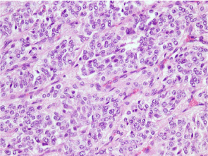
Figure 1
Cells arranged in nests (H&E, x400).
Figure 2
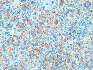
Figure 2
Tumor cells were positive for synaptophysin.
Table 1

Table 1
Immunoprofiles of thyroid paraganglioma and thyroid medullary carcinoma.
Discussion
Although the TP accounts for the 0.1 % of all the thyroid
neoplasms, this tumor must be remembered in the differential
diagnosis of the thyroid cancers [3].
The FNAB cannot be diagnostic in all cases preoperatively but
sometimes like in our case may give an idea about the suspicion of a
TP. Generally final pathology reports after the resection of the tumor
will give the definitive diagnosis. For the accurate diagnosis of the
pheochromocytomas and paragangliomas, both pathologists and
the clinicians must communicate properly. The importance of this
multidisciplinary approach has been popularized in recent studies.
Also an algorithm has been designed for the diagnosis of these cases
[6]. The paragangliomas can rarely be diagnosed by the radiologic
investigations and even diagnosis is difficult after the FNAB of the
nodule [7]. They can be easily be confused with the other thyroid
malignancies.
The diagnose frequently cannot be made during the surgery with
frozen sections and the differential diagnosis includes primarily the
medullary thyroid cancer and the other types of thyroid cancers.
Only 1 of the frozen sections performed in 22 patients made the
paraganglioma diagnosis. In 10 of these cases diagnosis of medullary
thyroid carcinoma and in 3 of the cases follicular carcinoma diagnosis were made by the frozen section [8,9].
The immunohistochemical stains applied has a major role in
the differential diagnosis [10,12-14]. The Chromogranin A, NSE,
Synaptophysin and S-100 will demonstrate the staining around the
Sustantacular cells. The S-100 staining however is not to specific
for the paragangliomas. It can be seen in the Medullary Thyroid
Cancer (MTC) too. Thus makes the diagnosis complex [10]. In this
case, paraganglioma presented as a mass located within the thyroid
gland. When present in such location, the main differential diagnosis
was MTC. On histologic examination, TP and MTC may show
similar microscopic features, with nesting. The co-expression of
neuroendocrine markers makes the distinction far more complicated.
MTC originates in the parafollicular C cells of the thyroid gland.
Therefore, MTC is characterized by positive staining with Calcitonin.
The positivity of epithelial markers and lack of S-100 positive
sustantecular cells also facilitate the differential diagnosis (Table 1)
[10,12-14].
They have a high tendency to invade (in two out of three cases)
adjacent structures like; trachea, recurrent nerves and vascular
structures. The extensive resection is generally enough for the
treatment of these patients [11]. The tumor was strictly attached to
the tracheal cartilages but any tracheal resection was not performed
in our patient.
The genetic mutations are reported in about %10 of the
paragangliomas. It is recommended to screen the patients for the
succinate dehydrogenase B (SDHB), C (SDHC), D (SDHD) or SDH
assembly factor 2 (SDHAF2) gene mutations. Especially it is known
that the SDHB mutation is important in diagnosing the malignant
paraganglioma and pheochromocytoma [8,9]. We did not perform a
genetic screening.
In the literature there is no reported case of metastatic or
catecholamine producing thyroid paraganglioma [8]. But still it
is advised to follow up those patients for the risk of metastasis and
catecholamine production [8]. Screening of the patient 29 months
after the surgery was performed and no metastasis or local recurrences
were seen in the ultrasound and PET scan (F-18 FDG). Serum and 24
hours urine tests were normal for the catecholamine and degradation
products.
Conclusion
TP must be considered in differential diagnosis of the neuroendocrine tumors of the thyroid. FNAB is helpful but not always diagnostic for a neuroendocrine tumor. Generally they are local invasive to the adjacent structures. Extensive resection and close follow up is the treatment of choice because their malignancy potential is still not exactly known. The long term post-operative follow up of these patients by the imaging modalities and the catecholamine levels is recommended.
References
- González Poggioli N, López Amado M, Pimentel MT. Paraganglioma of the thyroid gland: a rare entity. Endocr Pathol. 2009; 20: 62-65.
- Lack EE, Cubilla AL, Woodruff JM. Paragangliomas of the head and neck region. A pathologic study of tumors from 71 patients. Hum Pathol. 1979; 10: 191–218.
- Haegert DG, Wang NS, Farrer PA, Seemayer TA, Thelmo W. Nonchromaffin paragangliomatosis manifesting as a cold thyroid nodule. Am J Clin. Pathol. 1974; 61: 561–570.
- Phitayakorn R, Faquin W, Wei N, Barbesino G, Stephen AE. Thyroidassociated paragangliomas. Thyroid. 2011; 21: 725-733.
- Calò PG, Lai ML, Guaitoli E, Pisano G, Favoriti P, Nicolosi A, et al. Diffuculties in the diagnosis of thyroid paraganglioma: a clinical case. Clin Ter. 2013; 184: 35-39.
- Mete O, Tischler AS, de Krijger R, McNicol AM, Eisenhofer G, Pacak K, et al. Protocol for the examination of specimens from patients with pheochromocytomas and extra-adrenal paragangliomas. Arch Pathol Lab Med. 2014; 138: 182-188.
- Kafil Akhtar, Prasenjit Sen Ray, S Shamhsad Ahmad, Rana K Sherwani. Paraganglioma of the thyroid gland: cytologists' enigma. BMJ Case Rep. 2013; 22.
- Bao-Hua Yu, Wei-Qi Sheng, Jian Wang. Primary paraganglioma of thyroid gland: A clinicopathologic and immunohistochemical analysis of three cases with a review of the literature. Head and Neck Pathol. 2013; 7: 373-380.
- Stefan Costinean, Veronica Balatti, Arianna Bottoni, Mathew Old, Carlo Croce, Paul E. Wakely. Primary intrathyroideal paraganglioma: histopathology and novel molecular alterations. Hum Pathol. 2012; 43: 2371-2375.
- Erem C, Kocak M, Nuhoglu İ, Cobanoglu U, Ucuncu O, Okatan BK, et al. Primary thyroid paraganglioma presenting with double thyroid nodule: a case report. Endocrine. 2009; 36: 368-371.
- Armstrong MJ, Chiosea SI, Carty SE, Linwah Y. Thyroid paragangliomas are locally aggressive. Thyroid. 2012; 22: 88–93.
- Yano Y, Nagahama M, Sugino K, Ito K, Kameyama K, Ito K. Paranglioma of the thyroid: Report a male case with ultrasonographic imagings, cytologic, histologic, and immunohistochemical features. Thyroid. 2007; 17: 575-578.
- Esmeralda Castelblanco, Pilar Gallel, Susana Ros, Sonia Gatius, Joan Valls, Aguirre A. De-Cubas, et al. Thyroid paraganglioma. Report of 3 cases and description of an immunohistochemical profile useful in the differantial diagnosis with medullary thyroid carcinoma, based on complementary DNA array results. Hum Pathol. 2012: 43; 1103-1112.
- Bockhorn M, Sheu SY, Frilling A, Molmenti E, Schmid KW, Broelsch CE. Paraganglioma-like medullary thyroid carcinoma: a rare entity. Thyroid. 2005; 15: 1363-1367.