Case Report
Liver Metastasis from a Primary Mucinous Adenocarcinoma of the Ascending Colon with Metastatic Macroscopic Portal Vein Tumor Thrombus: A Case Report
Rubio DG*, Teh CS and Casupang A
National Kidney and Transplant Institute, Philippines
*Corresponding author: Donnel Guenter Rubio, National Kidney and Transplant Institute, East Ave, Diliman, Quezon City, Metro Manila, Philippines
Published: 23 Nov, 2016
Cite this article as: Rubio DG, Teh CS, Casupang A. Liver
Metastasis from a Primary Mucinous
Adenocarcinoma of the Ascending
Colon with Metastatic Macroscopic
Portal Vein Tumor Thrombus: A Case
Report. Ann Clin Case Rep. 2016; 1:
1187.
Abstract
We report a case of a metastatic macroscopic tumor thrombus of the portal vein arising from a primary colorectal cancer seen in one of the patients at the National Kidney and Transplant Institute, Philippines. The primary lesion is a mucinous adenocarcinoma of the ascending colon. Microscopic tumor thrombus from a colorectal cancer is quite rare compared to a tumor thrombus arising from a primary Hepatocellular carcinoma. Prognosis of patients having such tumor thrombus from a primary colorectal cancer is still unknown, but the primary surgical management is still liver resection with resection of tumor thrombus.
Introduction
Macroscopic portal vein tumor thrombus are usually seen in patients with primary hepatocellular carcinoma with an incidence rate of 22.5% on the other hand macroscopic portal vein tumor thrombus arising from a primary colorectal carcinoma is quite rare with an incidence of 2.8% and no case has yet to be reported in our country [1-2]. In this paper we present a case of a patient diagnosed to have mucinous adenocarcinoma of the ascending colon with a macroscopic portal vein thrombus on left portal vein positive for metastatic tumor cells.
Case Presentation
This is a case of S.W. a 30/F who was referred to our institution due to multiple liver masses on
segments 7, 8 4 and 5 seen thru whole abdominal CT scan. Her history started around december
2012 when the patient suddenly experienced abdominal pain crampy in character not relieved by
pain medications, not related to food intake radiating to right lower quadrant, she then consulted
a local hospital in their province, initial assessment during this time was ruptured appendicitis.
The patient then underwent explore laparotomy, intra operatively appendix was noted to be
congested, but there was an incidental finding of mid ascending colon mass, hence underwent right
hemicolectomy. Histopathology report of the mass showed a mucinous adenocarcinoma, eight of
thirteen lymph nodes positive for tumor. Further work ups were done, whole abdominal CT scan
was done and showed multiple masses on Segments 7,8,4 and 5 of the liver, largest on segment 711.6
x 1.5 x 1.5, segment 81.2 x 1.1x 1.5, segment 40.9 x 0.8 x 1.0, segment 50.9 x 0.7 x 0.6. Patient was
then referred to a medical oncologist and was started on oxaliplatin and xeloda. On repeat CT scan,
initially the liver masses responded to the treatment, but on surveillance CT scan done January
2015, there was a note of recurrence of the liver masses hence was referred to our institution.
On admission, repeat triphasic whole abdominal CT scan was done which showed hyperdense
mass in segment 6 and 7 of right lobe and a hypodense mass in segment 6 measuring 4.1 x 5.1 x 3.8
cm with associated dilatation of intrahepatic ducts in segments 6 and 7. Liver ultrasound was also
done which showed a solid heterogenous mass in segment 8 measuring approximately 6.4 x 6.0
x 5.4 cm. The mass has invaded and occluded the right portal vein. Patient then underwent right
hemihepatectomy with excision of nodules at segments 2 and 3 and left portal vein thrombectomy.
Intraoperatively there was a note of a 7 cm mass on segment 5 encroaching the right portal vein,
tumor thrombus noted at left portal vein and 1 cm nodules noted at segments 2 and 3 (Figure 1).
Post-operative histopathology
1. Right lobe: metastatic Colonic adenocarcinoma moderately differentiated with mucinous
features, resection margin negative for tumor.
2. Positive for tumor thrombus Potral vein.
3. Left portal thrombus - positive for tumor (metastatic).
Figure 1
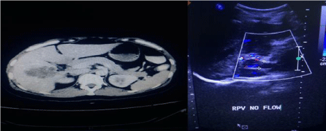
Figure 1
Right-whole abdominal CT scan showing liver mass. Left-liver
ultrasound with doppler showing the right portal vein.
Discussion
Tumor thrombus in the portal vein is common in patients having hepatocellular carcinoma. Tumor thrombus in the portal vein is seen in 64% of patients having HCCA. However metastatic lesions in the liver derived from colorectal cancer rarely invade the portal vein macroscopically, it is more associated with hepatocellular carcinoma. Colorectal liver metastases are usually accompanied by microscopic tumor invasion into the intrahepatic portal vein and incidence of macroscopic tumor thrombus in the portal vein is rare. Microscopic tumor invasion into the intrahepatic portal vein is detected in 20% of cases of liver metastases from colorectal cancer however for macroscopic tumor thrombus in portal vein is only 2.8%. In this paper we present a rare case of colorectal liver metastasis with portal vein tumor thrombus. As mentioned in a recent study done by Yamamoto et al. [3] most reported cases of portal vein tumor thrombus from colorectal cancer had concomitant metastatic nodules in the liver parenchyma as seen in this patient. Also mentioned in this study, macroscopic portal vein tumor thrombus form colorectal cancer displays a better prognosis compared to tumor thrombus arising from primary hepatocellular carcinoma, since presence of such in patients with HCCA would signify portal hypertension, rupture of esophageal varices or liver failure [4-6]. Despite presence or absence of portal vein tumor thrombus, best management for liver metastases is still surgical resection as what we did with this patient.
Conclusion
Patient had a successful resection of liver metastases from mucinous adenocarcinoma of ascending colon, with macroscopic tumor thormbus on left portal vein, although prognosis of such patients remains still unclear, resection of liver metastases with resection of tumor thrombus would appear to offer better prognosis.
References
- Yoshito Tomimaru, Yo Sasaki, Terumasa Yamada, Kunihito Gotoh, Shingo Noura, Hidetoshi Eguchi, et al. Liver metastasis originating from colorectal cancer with portal vein tumor thrombus. J Med Case Reports. 2010; 4: 382.
- Ogawa R, Kodama T, Kurishima K, Kagohashi K, Satoh H. Portal vein tumor thrombus of liver metastases from lung cancer. Acta Medica (Hradec Kralove). 2009; 52: 163-166.
- Naoto Yamamoto, Nobuhiro Sugano, Soichiro Morinaga, Amane Kanazawa, Daisuke Inagaki, Manabu Shiozawaet, et al. Massive portal vein thrombus from colorectal cancer without any metastatic nodules in liver parenchyma. Rare Tumors. 2011; 21; 3: 47.
- D Mathieu, N Vasile, P Grenier. Portal Thrombosis: dynamic Ct features and course. 1985; 154 3881793.
- Archi Agrawal, Nilendu Purandare, Sneha Shah, Ameya Puranik, Venkatesh Rangarajan. Extensive tumor thrombus of hepatocellular carcinoma in the entire portal venous system detected on PET scan. Indian J Nucl Med. 2013; 28: 54-56.
- Tsutomu Kobayashi, Hideki Suzuki, Norio Kubo, Akira Watanabe, Shigeru Sasaki, Wataru Wada, et al. A case of Hepatocellular carcinoma with portal vein tumor thrombosis successfully treated with intra arterial infusion of 5 Fluorouracil, Cisplatin and interferons. Int Surg. 2012; 97: 230–234.