Case Report
Intraparenchymal CSF Pseudocyst: An Unusual Complication of Ventriculoperitoneal Shunt
Alexander Lam* and Peter Y.C Gan
Department of Neurosurgery, Waikato Hospital, New Zealand
*Corresponding author: Alexander Lam, Department of Neurosurgery, Waikato Hospital, Hamilton 3204, New Zealand
Published: 23 Nov, 2016
Cite this article as: Lam A, Gan PYC. Intraparenchymal CSF Pseudocyst: An Unusual Complication of Ventriculoperitoneal Shunt. Ann Clin Case Rep. 2016; 1: 1186.
Abstract
Idiopathic Intracranial Hypertension (IIH) is most commonly seen in obese women between the ages of 20 and 44 years. Patients are initially managed conservatively with dietary modifications and medical treatment. Surgical intervention such asventriculoperitoneal shunt is usually reserved for patients with progressive deterioration of vision despite maximal medical therapy. Here we report a rare complication in a 25-year-old female who underwent ventriculoperitoneal shunt for IIH. Her symptoms improved initially following the procedure. However, she was readmitted a month later with acute worsening of symptoms and CT brain revealed a right occipito-parietal intraparenchymal cystic lesion of CSF density along the track of the ventricular catheter. The shunt was therefore removed and intraoperatively the peritoneal catheter was found to be obstructed. Culture of CSF and catheter samples were negative for infection. A new ventriculoperitoneal shunt was subsequently performed on the left side and her symptoms improved again immediately following surgery. To the best of our knowledge, this is the first reported case of an intraparenchymal CSF pseudocyst associated with ventriculoperitoneal shunt in the context of IIH and the third reported case of such complication in adult patients. We believe that the raised intracranial pressure and the blocked peritoneal catheter had synergistically resulted in the shunting of CSF from the ventricle into the interstitial space.
Introduction
ADC: Apparent Diffusion Coefficient; CSF: Cerebrospinal Fluid; CT: Computed Topography; DWI: Diffusion Weighted Imaging; IIH: Idiopathic Intracranial Hypertension; MRI: Magnetic Resonance Imaging
Introduction
Idiopathicintracranial Hypertension (IIH) is a neurological condition that comprises of signs and symptoms suggestive of a raised intracranial pressure in the absence of a space occupying lesion. It is therefore also known as pseudotumour cerebri. This condition is most commonly seen in obese women in their reproductive years. The incidence of IIH increases from 0.9/100,000 in the general population to 19.3/100,000 in women aged between 20 to 44 who weigh 20% or above their ideal body weight [1]. Patients with IIH typically complain of headache, blurry vision, diplopia and pulsatile tinnitus. On examination, impaired visual acuity, relative afferent pupillary defect, papilloedema, visual field defect and abducens nerve palsy are some of the frequent signs associated with this condition. The diagnostic criteria for IIH include: 1) Papilloedema; 2) Normal neurological examination other than cranial nerve deficits; 3) Normal neuroimaging to exclude space occupying lesions or cerebral venous sinus thrombosis; 4) Normal cerebrospinal fluid (CSF) composition and 5) A lumbar puncture opening pressure of 25 cm H2O or above [2]. The pathophysiology of this condition remains largely unclear and various aetiologies including interstitial cerebral oedema, increased CSF production, impaired CSF absorption andtransverse sinus stenosis have been proposed [3,4]. Initial treatment consists of dietary modifications to induce weight loss and medications such as acetazolamide and frusemide to reduce CSF production. Surgical interventions including ventriculoperitoneal shunt and lumboperitoneal shunt are usually reserved for patients with progressive deterioration of vision despite maximal medical treatment. Here we report an unusual complication associated with ventriculoperitoneal shunt in a patient with IIH.
Case Presentation
R D is a 25 year-old female on a background history of polycystic ovarian syndrome and morbid obesity with a body mass index of 48.5. She presented with two weeks history of worsening generalised headache associated with blurry vision, memory impairment and sense of imbalance. On examination, she had 6/6 visual acuity in both eyes with no visual field defect to confrontation or relative afferent pupillary defect. Fundoscopy revealed bilateral papilloedema and the rest of her neurological examination was otherwise unremarkable. Initial contrast enhanced CT brain showed no evidence of any space occupying lesions or venous sinus thrombosis (Figure 1). Under fluoroscopic guidance, lumbar puncture was performed and the opening pressure was noted to be at least 60 cm of H2O. The exact pressure was unable to be measured as the opening pressure exceeded the maximal scale of the manometer. Following drainage of 42 ml of CSF, the closing pressure reduced to 6 cm of H2O. The diagnosis of IIHwas made and she was commenced on acetazolamide. Her symptoms including headache and blurry vision however continued to deteriorate despite maximal medical treatment with the primary intention to preserve her vision, she was consented for the insertion of a right ventriculoperitoneal shunt with a programmable valve during the same admission (Figure 2). Her symptoms improved significantly following surgery, acetazolamide was ceased and she was subsequently discharged home. One month following her initial surgery, she returned with acute deterioration of headache and blurry vision. Her visual acuity deteriorated from 6/6 to 6/24 bilaterally with fundoscopy revealing evidence of papilloedema in both eyes. CT brain revealed a 31mm x 39mm x 33mm intraparenchymal cystic lesionof CSF density along the track of the ventricular catheter in the right occipito-parietal region (Figure 3). The tip of the ventricular catheter remained in the right lateral ventricle and no contrast enhancement was seen in this cystic lesion or adjacent parenchymal tissue. The ventriculoperitoneal shunt was removed due to the concern of shunt malfunction and infection and she was commenced on empirical antibiotics. Intraoperatively the ventricular catheter was noted to be draining CSF under moderate pressure and the intracranial pressure was measured to be 31 cm of H2O via the ventricular catheter. The peritoneal catheter appeared to be obstructed distally with evidence of retrograde flow. Cultures from CSF and catheters were negative for bacterial or fungal infection. Following the removal of the ventriculoperitoneal shunt, a MRI scan was performed to further investigate this intraparenchymal cystic lesion. On MRI, this cystic lesion appeared to be T1 hypointense and T2 hyperintense with no enhancement with gadolinium (Figure 4). It was associated with oedema in the adjacent parenchyma and no diffusion restriction was seen on diffuse weighted imaging (DWI) and apparent diffusion coefficient (ADC) to suggest ischaemic stroke. Considering the progressive worsening of papilloedema and the clinical suspicion for infection is reasonably low, a left sided ventriculoperitoneal shunt was inserted on the next day following the removal of the right sided shunt. Fenestration of the left optic nerve sheath was performed by the ophthalmologist during the same procedure. Her headache and blurry vision improved significantly following the procedure. Post-operative CT brain showed significant reduction in the size of this cystic lesion (Figure 5A). At one-month follow up, her visual acuity with pinhole improved to 6/6 on the right and 6/9 on the left with 100% red saturation bilaterally and residual relative afferent pupillary defect in the left eye. Fundoscopy revealed mild residual papilloedema in bilateral nasal discs. Confrontational visual fields to red target were intact. At three-month follow up, her visual acuity continued to improve to 6/5 on the right and 6/6 on the left unaided with no evidence of relative afferent pupillary defect. Progress CT brain at eight-month follow up demonstrated further regression of the cystic lesion (Figure 5B).
Figure 1
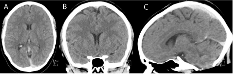
Figure 1
Initial CT brain in axial (A), coronal (B) and sagittal (C) sections demonstrating no evidence of space occupying lesions.
Figure 2
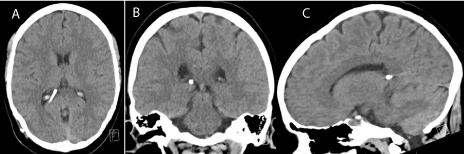
Figure 2
CT brain following the insertion of right ventriculoperitoneal shunt, demonstrating the tip of the ventricular catheter in the right lateral ventricle in axial (A), coronal (B) and sagittal (C) sections.
Figure 3
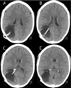
Figure 3
CT Brain one month following the insertion of right
ventriculoperitoneal shunt, demonstrating the intraparenchymal cystic lesion
of CSF density in the right occipito-parietal region in axial sections in a
cranio-caudal order from A to D.
Discussion
Based on the radiological features of the lesion and our intraoperative findings, we believe that this lesion is most consistent with an intraparenchymal CSF pseudocyst. The ventricular catheter we used in this case was an antibiotic impregnated Bactiseal® EVD catheters (Codman Neuro DePuy Synthes). This catheter consists of fenestrations in the initial 3 cm segment from the tip. Even though the tip of the ventricular catheter was located in the right lateral ventricle as shown on the post-operative CT scan, a portion of the fenestrated segment could still be located in the brain parenchyma. As the peritoneal catheter became obstructed distally, the intracranial pressure began to rise and eventually the resultant hydrostatic pressure became so high that it forced CSF to flow from the ventricular system into the interstitial space to form a pericatheter CSF pseudocyst. It is considered as a pseudocyst as it lacks an epithelialised wall. There were two possible pathways that would allow for the shunting of CSF from the ventricle into the interstitium. One wasvia the fenestrations in the distal segment of the ventricular catheter and there other one was through the track along the catheter that had been created during its insertion. As the CSF cyst continued to expand, it behaved as aspace-occupying lesion and contributed to a further increase in the intracranial pressure. Since the underlying cause of the intraparenchymal CSF pseudocyst was shunt malfunctioning, it was effectively managed with revision of the ventriculoperitoneal shunt as demonstrated in the follow up CT scans in this case. Other differential diagnoses such as infection and ischaemic stroke were also considered. However, infection was deemed less possible given the absence of infective symptoms, the lack of raised inflammatory markers and the negative cultures from CSF and catheter samples. Similarly, there was no evidence of diffusion restriction on MRI to suggest ischaemic stroke. Several cases of ventriculoperitoneal shunt associated CSF pseudocysts have been reported previously with a majority of these cases were seen in paediatric patients [5-8] and only two cases were reported in adult patients [9,10]. Therefore, to the best of our knowledge, this is the third reported case in adult. Furthermore, this is also the first reported case of a shunt related pericatheter CSF pseudocyst in the context of IIH.
Figure 4
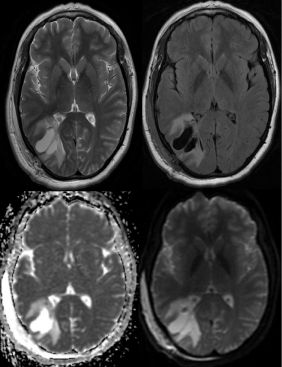
Figure 4
MRI brain following the removal of the right ventriculoperitoneal
shunt in T2 sequence (A), FLAIR sequence (B), DWI (C) and ADC (D). This
cystic lesion appeared to be T2 hyperintense associated with oedema in
surround tissue. While it is of high signal intensity in DWI, ADC shows no
corresponding low signal intensity to suggest true diffusion restriction.
Figure 5
Figure 5
CT Brain following the insertion of left ventriculoperitoneal shunt
on post-operative day 5 (A) and at 8 month follow up (B), demonstrating a
gradual resolution of the right occipito-parietal cystic lesion.
Conclusion
As the global obesity epidemic continues to worsen, the number of ventriculoperitoneal shunt that is performed for IIH is also likely to increase in the near future. We therefore hope to raise the awareness of such a rare yet possible complicationamongst clinicians.
References
- Durcan FJ, Corbett JJ, Wall M. The incidence of pseudotumor cerebri. Population studies in Iowa and Louisiana. Arch Neurol. 1988; 45: 875-877.
- Friedman DI. The pseudotumor cerebri syndrome. Neurol Clin. 2014; 32: 363-396.
- Joynt RJ, Sahs AL. Brain swelling of unknown cause. Neurology. 1956; 6: 801-803.
- Quattrone A, Bono F, Oliveri RL, Gambardella A, Pirritano D, Labate A, et al. Cerebral venous thrombosis and isolated intracranial hypertension without papilledema in CDH. Neurology. 2001; 57: 31-36.
- Iqbal J, Hassounah M, Sheikh B. Intraparenchymal pericatheter cyst. A rare complication of ventriculoperitoneal shunt for hydrocephalus. Br J Neurosurg. 2000; 14: 255-258.
- Sinha AK, Lall R, Benson R, O'Brien DF, Buxton N. Intraparenchymal pericatheter cyst following ventriculoperitoneal shunt insertion: does it always merit shunt revision?. Zentralbl Neurochir. 2008; 69: 152-154.
- Rim HR, Hwang SK, Kwon SH, Kim HM. Intraparenchymal pericatheter cyst as a complication of a ventriculo-peritoneal shunt in a premature infant. J Korean Neurosurg Soc. 2011; 50:143-146.
- Sugimoto K, Enomoto T, Nose T. Reversible porencephaly. Alteration of the cerebrospinal fluid flow after shunt malfunction. Childs Nerv Syst. 1991; 7: 394-398.
- Vajramani GV, Fugleholm K. Reversible CSF cyst related to a functioning ventriculo-peritoneal shunt. Acta Neurochir (Wien). 2005; 147:1199-1202.
- Amans MR, Dillon WP. Cerebral parenchymal cyst: A rare complication of ventriculoperitoneal shunt malfunction in an adult. Radiol Case Rep. 2013; 8: 784.