Case Report
Malignant Transformation of a Teratoma Presenting with Metastasis to the Hip
McCready Mariah* and Jozef Malysz
Department of Pathology, Penn State Milton S Hershey Medical Center, USA
*Corresponding author: McCready Mariah, Department of Pathology, Penn State Milton S Hershey Medical Center, 500 University Drive, Hershey, Pa 17033, USA
Published: 23 Nov, 2016
Cite this article as: McCready Mariah, Malysz J. Malignant
Transformation of a Teratoma
Presenting with Metastasis to the Hip.
Ann Clin Case Rep. 2016; 1: 1184.
Abstract
Teratomas are germ cell tumors composed of cells recapitulating the three germ cell layers. Malignant transformation of a teratoma (TMT) is defined as a non-germ cell malignant tumor arising in a preexisting mature teratoma, and it is a rare occurrence. Metastases associated with malignant transformation are even more infrequent, mostly having been reported arising from gonadal teratomas. This report describes a case of a 41 year old female presenting with metastatic adenocarcinoma to the hip as the initial clinical manifestation of malignant transformation of a mediastinal teratoma, along with a review of relevant literature.
Introduction
Teratomas are germ cell tumors composed of cells recapitulating the three germ cell layers. They are most common in the gonads; however they can occur any where along the midline axis due to
migration of germ cells during embryogenesis. Teratomas are the most common germ cell tumor
of the mediastinum [1]. Malignant transformation of a teratoma (TMT) is defined as a non-germ
cell malignant tumor arising in a preexisting mature teratoma, and it is a rare occurrence. The nongerm
cell component can be a carcinoma or sarcoma. This is to be differentiated from malignant
teratoma, which includes TMT, as well as teratoma with a malignant germ cell component, and a
combination of germ cell and non-germ cell components [2].
Metastases associated with malignant transformation are even more infrequent, mostly
having been reported arising from gonadal teratomas. This report describes a case of metastatic
adenocarcinoma to the hip as the presenting manifestation of malignant transformation of a
mediastinal teratoma.
Case Presentation
A 41-year old female presented with several months of right hip pain following a fall. There
was no improvement with medications and physical therapy, and so the patient was evaluated by
orthopedics. She was found to have an expansile lesion in the right femur, with impending fracture, as
well as multiple other areas suspicious for metastatic disease. She under went an orthopedic pinning
procedure, and a biopsy taken at the time of surgery demonstrated a metastatic adenocarcinoma
of nonspecific phenotype (Figure 1). Immunohistochemical stains revealed the tumor to highlight with chromogen against pankeratin, polyclonal CEA, and CK 20. The neoplastic cells werenegative
for CK7, as well as TTF-1, CDX2, GCDFP, ER, PR, CA19.9, CA125, and p63. These results were
considered non-specific with respect to a possible primary site.
Subsequent imaging demonstrated an anterior mediastinal mass. The resection of the mass
was a 4.5x2.5x2 cm unilocular cyst containing hair and sebaceous material. The cyst wall ranged in
thickness from 0.1 to 1.0 cm, and areas of the lesion were completely solid, consisting of tan-white
soft tissue. Microscopic examination revealed the presence of bone, smooth muscle, hair, respiratory
epithelium, and intestinal epithelium, consistent with a teratoma. Within the intestinal epithelium,
there were dysplastic changes, which ranged from low grade, as in a tubular adenoma, to high grade,
with loss of nuclear polarity and increased nuclear pleomorphism. This latter area was associated
with invasive glands present in the wall of the tumor, consistent with invasive adenocarcinoma
(Figure 2). The majority of the adenocarcinoma was poorly differentiated, growing in cords, sheets, and single cell pattern. Immunohistochemical stains on the mediastinal mass revealed the tumor
was positive for CK20 and CDX2 and negative for CK7 and TTF-1. Given the presence of an insitu
component to the tumor, as well as the immunohistochemical staining pattern, the tumor was
diagnosed as an invasive adenocarcinoma of intestinal type, arising in a mature teratoma.
The patient received chemotherapy and radiation for her
metastatic disease. The last report available had her doing well at a 2
month post-operatively. The patient was subsequently lost to followup.
Figure 1
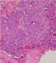
Figure 1
Poorly differentiated adenocarcinoma in right femur curettings.
Figure 2
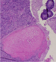
Figure 2
Mediastinal mass with mesenchymal tissue, adenomatous tissue,
and infiltrative carcinoma.
Discussion
TMT is a rare occurrence; present in 1-2% of ovarian mature
teratomas [3]. The incidence in mediastinal teratomas has not been
widely studied, however one large study of 322 cases of primary
mediastinal germ cell tumors found it to occur in 4% of teratomatous
lesions in this site [2]. The majority of cases of TMT have been reported
in men [4,5]. A study of 17 male patients found that TMT localized
to the testis had a better prognosis than those with disease outside the
testis, including the mediastinum [5]. Several studies have found that
the most common malignant non-germ cell differentiation in men
with TMT was sarcomatous, followed by carcinomatous; the usual
types of sarcoma included rhabdomyosarcoma, undifferentiated
sarcoma, primitive neuroectodermal tumor, chondrosarcoma, and
malignant peripheral nerve sheath tumor [5-8]. In contrast, the
majority of females with ovarian TMT show an epithelial malignancy,
with squamous cell carcinoma being the most common diagnosis [3].
Four cases of metastatic disease arising from a mediastinal TMT
have been reported previously. As opposed to the current case,
the metastasis in these cases were found after the primary tumor
had been diagnosed or in conjunction with the primary tumor.
The metastatic tumor in these cases was sarcoma in two cases and
adenocarcinoma in the other half [9-12]. One paper describes that
half of the patients with malignant teratomas have metastasis outside
of the mediastinum, however it does not distinguish between the
different types of malignant teratoma, and so the true incidence of
metastatic disease in mediastinal TMT is still not known [13,14].
To the best of our knowledge, this is the first reported case of
a metastasis as the presenting manifestation of a TMT arising in
the mediastinum. In addition, this case is in a female, which is not
widely reported in the literature. This case illustrates the need to keep
a wide differential when faced with a metastatic carcinoma with an
unknown primary. Had the metastatic carcinoma showed the same
immunohistochemical profile as the primary tumor (CK 20 and
CDX2 positive, CK7 negative), the likely interpretation would have
been that of a gastrointestinal, probably colonic, primary malignancy.
This could have led down the wrong diagnostic pathway, and
demonstrates a possible pitfall of depending on immunohistochemical
stains to determine the primary site of a metastatic carcinoma. It is
well known that the decalcification process can lead to false results
with immunohistochemical staining. We submit the proposal that
after the diagnosis of metastatic tumor, especially adenocarcinoma, is
made, a thorough work-up, by imaging and blood work, may be the
best way to discover the primary site. While immunohistochemical
stains can be instrumental in narrowing down the possible primary
sites of a metastatic tumor, care should be taken to not exclude other
possibilities based on their results.
References
- Nichols CR. Mediastinal germ cell tumors: clinical features and biologic correlates. Chest. 1991; 99: 472-479.
- Moran CA, Suster S. Primary germ cell tumors of the mediastinum: analysis of 322 cases with special emphasis on teratomatous lesions and a proposal for histopathologic classification and clinical staging. Cancer. 1997; 80: 681-690.
- Chadha S, Schaberg A. Malignant transformation in benign cystic teratomas; dermoids of the ovary. Eur J Obstet Gynecol Reprod Bio. 1988; 29: 329-398.
- Ulbright TM, Loehrer PJ, Roth LM, Einhorn LH, Williams SG, Clark SA. The development of non-germ cell malignancies within germ cell tumors: a clinicopathologic study of 11 cases. Cancer. 1984; 54: 1824-1833.
- Ahmed T, Bosl GJ, Hajdu SI. Teratoma with malignant transformation in germ cell tumors in men. Cancer. 1985; 56: 860-863.
- Comiter CV, Kibel AS, Richie JP, Nucci MR, Renshaw AA. Prognostic features of teratomas with malignant transformation: a clinicopathological study of 21 cases. J Urol. 1998; 159: 859-863.
- Athanasiou A, Vanel D, El Mesbahi O, Theodore C, Fizazi K. Non-germ cell tumours arising in germ cell tumours (teratoma with malignant transformation) in men: CT and Mr findings. Eur j Rad. 2009; 69: 230-235.
- Motzer RJ, Amsterdam A, Prieto V, Sheinfeld J, Murty VVVS, Mazumdar M, et al. Teratoma with malignant transformation: diverse malignant histologies arising in men with germ cell tumors. J Urol. 1998; 159: 133- 138.
- Duran I, Riveros L, Berthold D, Sweet J, Moore M. Eyelid metastases from mediastinal teratoma with malignant transformation. Acta Oncol. 2007; 46: 1200-1201.
- Popp G, Dragnev K. Secondary malignant transformation of a primary mediastinal germ cell tumor with diffuse lymphangitic spread to the lungs. South Med J. 2003; 96: 696-698.
- Kim HJ, Kim HR. Naturally occurring Mediastinal Teratoma with Malignant Transformation in an Adult Male. Korean J Thoracic Cardiovascular Surg. 2013; 46: 305-308.
- Templeton, A. Malignant Mediastinal Teratoma with Bone Metastases. Radiology. 1961: 245-247.
- Wychuliz AR, Payne WS, Clagett OT, Woolner LB. Surgical treatment of mediastinal tumors: a forty-year experience. J Thorac Cardiovasc Surg. 1971; 62: 379-392.