Case Report
Cervicofacial Emphysema After Routine Dental Procedures: An Iatrogenic Complication or Odontogenic Infection with Necrotizing Fasciitis?
Aditi Mohankumar*, Nnenna Ezeilo and Carol A Bauer
Department of Otolaryngology, Southern Illinois University School of Medicine, USA
*Corresponding author: Aditi Mohankumar, Department of Otolaryngology, Southern Illinois University School of Medicine, P.O. Box 19662, Springfield, IL 62794, USA
Published: 12 Nov, 2016
Cite this article as: Mohankumar A, Ezeilo N, Bauer CA.
Cervicofacial Emphysema After Routine
Dental Procedures: An Iatrogenic
Complication or Odontogenic Infection
with Necrotizing Fasciitis?. Ann Clin
Case Rep. 2016; 1: 1181.
Abstract
A number of both infectious and non-infectious etiologies may result in cervicofacial emphysema. Iatrogenic subcutaneous emphysema occurring afterdental instrumentation is usually a benign and self-limited process. However, it remains critical to rule out ominous causes of cervicofacial emphysema, namely necrotizing fasciitis. Here, we discuss the case of an individual with a history of recent dental procedures presenting with clinical and radiologic findings concerning for necrotizing fasciitis. Subsequent neck exploration was unremarkable, indicating the preceding dental instrumentation as the likely iatrogenic source of his extensive cervical subcutaneous emphysema. Therefore, we also discuss strategies to help differentiate necrotizing fasciitis from other benign causes of cervical emphysema.
Keywords: Prucalopride; Varenicline; Serotonin; Receptors; Interaction; Case report
Introduction
Cervicofacialemphysema, defined as the abnormal presence of air within the soft tissue of the
head and neck, may result from a number infectious and non-infectious etiologies. This abnormal
introduction of air into soft tissues then propagates along interconnected fascial planes [1,2]. Known non-infectious causes include blunt trauma [3], airway foreign bodies [4,5], abdominal
surgery [6,7], tracheal injury [8], and spontaneous rupture of terminal alveoli [9]; while infectious
causes include odontogenic infection [10] and necrotizing fasciitis. Cervicofacial emphysema
was first reported in 1900, when an individual developedremarkable cervicofacial swelling while
playing the bugle after a dental extraction [11]. Since then, various dental interventions including
dental extraction, endodontic treatment, and restorative dentistry have been associated with
subcutaneous emphysema [1,2]. Historically, patient actions such as coughing, vomiting, or other valsalva maneuvers following dental extraction preceded cervicofacial emphysema. Presently, this
complication commonly occurs after the use of sophisticated air-driven hand-pieces and high-speed
drilling dental instruments [2,12].
With few exceptions, cervicofacial emphysema resulting from dental procedures is a relatively
benign and self-limited process. Patients are often managed conservatively with rare need for
surgical intervention [11]. In certain situations, the clinical presentation may be mistaken for the
emergent condition of cervicofacial necrotizing fasciitis [NF]. NF is a rare, progressive infection
with necrosis of soft tissue, subcutaneous fat and skin. NF has an incidence on 0.40 cases per 100,000
people [13]. Even much rarer, cervicofacial necrotizing fasciitis represents about 5% of NF cases and is usually caused by dental infection, oropharyngeal infection, and infection secondary to trauma
[13,14].
A diagnostic conundrum exists in early NF since non specific clinical features such as erythema,
tenderness, warm skin and swelling are present. Therefore, identifying the underlying cause of
cervicofacial emphysema is central to appropriate and judicious management of these patients.
Head and neck subcutaneous emphysema after dental procedures have been extensively discussed in
the journals of dental medicine, while this complication has been rarely reported in otolaryngology
literature despite its pertinent to the field [1]. Here, we present the case of a diabetic, ill-appearing 32-year-old male with cervicofacial emphysema of unclear etiology. At initial presentation, this
was suspected to be an odontogenic infection after a dental extraction progressing to necrotizing
fasciitis. We also discuss strategies to facilitate the critical differentiation of necrotizing fasciitis from
benign causes of cervical emphysema.
Case Presentation
A 32-year-old male with a recent history of a dental procedures,
presented to the Emergency Department with complaint of right
jaw pain, severe nausea with emesis and malaise. He had a history
of right mandibular premolar tooth extraction without incidence
two weeks ago prior to presentation and a routine dental cleaning
on the day of presentation. His medical history was significant for
type 2 diabetes mellitus. On evaluation, the patient’s vital signs were
as follows: afebrile with a temperature 36.6ºC, tachycardia with heart
rate of 108 beats per minute and hypertension with blood pressure of
140/91 mmHg. Abnormal blood tests include leukocytosis with white
blood count of 21.1 k/cumm with 95% neutrophils and blood glucose
level of 265 mg/dL. Physical examination was significant for right
mandibular angle tenderness, tooth socket with fibrinous exudate
and mild crepitus along right mandible; there was also mild erythema
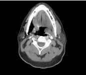
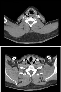
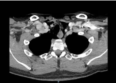
of anterior neck and supraclavicular area. A CT soft tissue neck
demonstrated extensive cervical emphysema within the soft tissue of
the neck centered on the right mandible and extending inferiorly to
the mediastinum (Figures 1-3). No obvious fluid collections noted on
imaging. Flexible fiberoptic laryngoscopy was unremarkable without
any laryngeal edema or erythema.
The constellation of clinical findings was concerning for
odontogenic infection with progression to cervicofacial necrotizing
fasciitis. After the initiation of broad spectrum parenteral antibiotics
with vancomycin and piperacillin/tazobactam, the patient was
emergently taken to the operating room for a neck exploration and
debridement. This neck exploration was negative for necrotic tissue
or purulence along the deep fascial planes, with presence of healthyappearing
bleeding tissue noted. A wound swab was obtained from
the neck as well as tooth socket. Normal flora was noted on the
wound culture from the neck, while the culture from the tooth
socket showed normal oral flora including moderate non-influenzae
Haemophilus species. Since necrotizing fasciitis was eliminated,
the patient’s preceding dental cleaning on the day of presentation
was surmised to be the likely cause of his cervicofacial emphysema
and pneumomediastinum. It is suspected that a compressed airdriven
instrument such as a tooth dryer may have been used during
this dental cleaning. The patient had a four-day hospital stay, with
normalization of his white blood cell count to 9.5 and transition to
oral Augmentin at discharge.Cervical erythema and crepitus had
resolved prior to discharge.
Figure 1
Figure 1
Axial CT neck demonstrates subcutaneous emphysema around
the right mandible and muscles of mastication.
Figure 2
Figure 2
Axial CT neck shows subcutaneous emphysema surrounding
the carotid sheath, cervical trachea, and dissecting the retropharyngeal/
retroesophageal space.
Figure 3
Figure 3
Axial CT chest demonstrates pneumomediastinum with
subcutaneous air around the intrathoracic trachea and esophagus.
Discussion
Cervicofacial emphysema resulting from dental procedures
represents a relatively benign and self-limited process. A variety
of compressed air-driven hand-piece instruments used in dental
interventions have been implicated, and have been used in different
procedures ranging from endodontics, restorative procedures, crown
preparation to dental extractions [1,2,12]. For instance, high-speed
air turbine drills designed for cutting teethare driven by compressed
air at 3.5 to 4.0 Newton-meter, and can rotate at 450,000 revolutions
per minute [15]. Air may be directed towards the burr to act as
a coolant, which can result in the introduction of air at vulnerable
sites. Similarly, a number of canal cleansing devices have been
shown to generate positive apical pressures greater than of central
venous pressure [16-19]. In most cases subcutaneous emphysema,
patients can be conservatively managed with close observation and
intravenous antibiotics [2,20,21]. Symptoms typically resolve within several days with no long-term sequelae.
However, it is imperative to distinguish an iatrogenic
complication from more ominous causes of cervicofacial emphysema,
namely necrotizing fasciitis [NF]. Cervicofacial NF can occur from
oropharyngeal or odontogenic infection, often in the setting of
a predisposing systemic illness, commonly diabetes mellitus or
immunodeficiency [14,22,23]. Clinical features of early NF such
as pain, swelling and erythema of affected area are subtle, nonspecific
and easily confused with other skin and soft tissue infections
[23,13,24]. Findings such as blistering, necrosis, and crepitus typically
occur later in the disease process, and may still be present only in 10-
40% of patients [13]. This may lead to missed or delayed diagnosis
in the early stages of this disease. Lancerotto et al. [13] report that
85-100% of these patients may have missed or delayed diagnosis at
presentation, which resulted in significantly worse outcomes with
increased mortality rates. In their retrospective review, Wong et al.
[25] found that only 13 out of 89 patients with confirmed necrotizing
fasciitis were appropriately diagnosed at admission. The authors
also noted that delay in surgery of greater than 24 hours correlated
with significantly increased mortality rates (RR = 9.4, p <0.05) [25].
Unsurprisingly, mortality is significantly decreased in patients with
early aggressive surgical debridement when compared to delayed or
incomplete surgical debridement (4.2% vs. 38%, p = 0.0007) [26].
In order to facilitate accurate and prompt diagnosis of NF, several
evaluation tests including imaging, a scoring system and bedside
biopsy have been described [23,27]. Relevant findings on computed
topography and magnetic resonance imagingare thickening and
enhancement of the skin, subcutaneous tissue, fascia, and muscle [28],
with lack of facial enhancement on contrast administration suggested
to be of paramount diagnostic value [29,30]. The presence of air on
imaging is variable, reported in 16.9% - 83% of patients, and may not
be specific to necrotizing fasciitis [23,25,29]. In 2004, Wong et al.
[30] proposed the Laboratory Risk Indicator for Necrotizing Fasciitis
(LRINEC) score, which uses serologic values to stratify patients into
three risk groups: low, intermediate and high risk groups. LRINEC
scores is estimated using: the serum levels of C-reactive protein, white
blood count, creatinine, glucose, and sodium. ALRINEC score ≥6
carries a greater than 50% risk of NF, therefore NF must be carefully
ruled out. Notably, the LRINEC score does not establish a definitive
diagnosis and can be elevated in non-necrotizing severe soft tissue
infections [31-34].
In an effort to prevent treatment delays, several adjunctive bedside
procedures to facilitate diagnosis of NF have been described. Some
authors report performing a small incision and wound exploration
at bedside [35,36]. In NF, grossly necrotic tissue, absence of bleeding,
fascial edema, and thin foul-smelling pus would be noted. Andreasen
et al. [36] also suggest that minimal resistance with finger dissection
of the deep cervical fascia can help confirm this diagnosis. Bedside
tissue biopsy with frozen section analysis may also be beneficial
[36-38]. In their case series of NF patient over 15 years, Majeksi &
Majeski used a bedside biopsy to accurately diagnosis NF in 12 of 43
patients, and NF was confirmed on subsequent surgical exploration;
the remaining 31 patient had non-NF infectious process such as
cellulitis or abscess [38]. Under local anesthesia with 1% lidocaine,
the authors described obtaining an elliptical biopsy 2cm x 1cm of skin
with underlying soft tissue & fascia of the affected area. This tissue
biopsy is immediately taken for gram stain and frozen section, with
their pathologist establishing a diagnosis of NF within 15 minutes of receiving the specimen. However, surgical exploration remains the most sensitive and reliable diagnostic tool to confirm or exclude NF [13].
This case illustrates that cervicofacial emphysema can have a
broad non-infectious differential. Subcutaneous emphysema can
occur as an iatrogenic complication of dental procedures when airdriven
hand-piece dental instruments are used. Non-infectious
cervicofacial emphysema is often a self-limited process that
improves with conservative management, but can be mistaken for
early NF given nonspecific clinical features. In the case presented,
the constellation of clinical findings coupled with the cervicofacial
emphysema in this patient strongly suggested early necrotizing
fasciitis from an odontogenic infection after a dental extraction: the
patient had a systemic inflammatory response with chills, tachycardia,
malaise, and was toxic-appearing, in addition to his poorly controlled
diabetes mellitus conferring an immunocompromised state. The high
clinical suspicion for necrotizing fasciitis warranted early surgical
exploration in this patient, since clinical and radiology findings are
often non-specific in early NF [13] and delay in surgical exploration
is associated with increased risk of mortality in NF [23,25]. A serum
C-reactive protein was not obtained and thus a LRINEC score could
be calculated in this case. None the less, the authors concede that
a limited bedside biopsy could have been used as an intermediate
procedure to further guide clinical decision making. A “watch and
wait” approach with parenteral antibiotics is potentially risky if NF
is suspected given high mortality rates, ranging from 19-40%. When
presentation is equivocal, consider the use of adjunctive bedside
procedures such as incision with wound exploration or tissue biopsy
with frozen section analysis, to help confirm or exclude the diagnosis
of necrotizing fasciitis.
References
- An GK, Zats B, Kunin M. Orbital, mediastinal and cervicofacial subcutaneous emphysema after endodontic retreatment of a mandibular premolar: a case report. J Endod. 2014; 40: 880-883.
- Heyman SN, Babayof I. Emphysematous complications in dentistry, 1960- 1993: an illustrative case and review of the literature. Quintessence Int. 1995; 26: 535-543.
- Chouliaras K, Bench E, Talving P, Strumwasser A, Benjamin E, Lam L, et al. Pneumomediastinum following blunt trauma: Worth an exhaustive workup? J Trauma Acute Care Surg. 2015; 79: 188-192.
- Hu M, Green R, Gungor A. Pneumomediastinum and subcutaneous emphysema from bronchial foreign body aspiration. Am J Otolaryngol. 2013; 34: 85-88.
- Kumar M, Goyal A, Gupta N, Rautela RS. Subcutaneous emphysema: Unique presentation of a foreign body in the airway. J Anaesthesiol Clin Pharmacol. 2015; 31: 404-406.
- Jones A, Pisano U, Elsobky S, Watson AJM. Grossly delayed massive subcutaneous emphysema following laparoscopic left hemicolectomy: A case report. Int J Surg Case Rep. 2015; 6: 277-279.
- Montori G, Di Giovanni G, Mzoughi Z, Angot C, Al Samman S, Solaini L, et al. Pneumoretroperitoneum and Pneumomediastinum Revealing a Left Colon Perforation. Int Surg. 2015; 100: 984-988.
- Barrett E. Management of a traumatic tracheal tear: a case report. AANA J. 2011; 79: 468-470.
- Parker GS, Mosborg DA, Foley RW, Stiernberg CM. Spontaneous cervical and mediastinal emphysema. Laryngoscope. 1990; 100: 938-940.
- Steiner M, Grau MJ, Wilson DL, Snow NJ. Odontogenic infection leading to cervical emphysema and fatal mediastinitis. J Oral Maxillofac Surg. 1982; 40: 600-604.
- Turnbull A. A Remarkable Coincidence in Dental Surgery. Br Med J. 1900; 1: 1131.
- McKenzie WS, Rosenberg M. Iatrogenic subcutaneous emphysema of dental and surgical origin: a literature review. J Oral Maxillofac Surg. 2009; 67: 1265-1268.
- Lancerotto L, Tocco I, Salmaso R, Vindigni V, Bassetto F. Necrotizing fasciitis: classification, diagnosis, and management. J Trauma Acute Care Surg. 2012; 72: 560-566.
- Yadav S, Verma A, Sachdeva A. Facial necrotizing fasciitis from an odontogenic infection. Oral Surg Oral Med Oral Pathol Oral Radiol. 2012; 113: 1-4.
- Arai I, Aoki T, Yamazaki H, Ota Y, Kaneko A. Pneumomediastinum and subcutaneous emphysema after dental extraction detected incidentally by regular medical checkup: a case report. Oral Surg Oral Med Oral Pathol Oral Radiol Endod. 2009; 107: 33-38.
- Khan S, Niu LN, Eid AA, Looney SW, Didato A, Roberts S, et al. Periapical pressures developed by nonbinding irrigation needles at various irrigation delivery rates. J Endod. 2013; 39: 529-533.
- Eleazer PD, Eleazer KR. Air pressures developed beyond the apex from drying root canals with pressurized air. J Endod. 1998; 24: 833-836.
- Durukan P, Salt O, Ozkan S, Durukan B, Kavalci C. Cervicofacial emphysema and pneumomediastinum after a high-speed air drill endodontic treatment procedure. Am J Emerg Med. 2012; 30: 2095-2099.
- Gulati A, Baldwin A, Intosh IM, Krishnan A. Pneumomediastinum, bilateral pneumothorax, pleural effusion, and surgical emphysema after routine apicectomy caused by vomiting. Br J Oral Maxillofac Surg. 2008; 46:136-137.
- Lococo F, Trabucco L, Leuzzi G, Salvo F, Paci M, Sgarbi G, et al. Severe breathing and swallowing difficulties during routine restorative dentistry. Ann Ital Chir. 2015; 86: 1-3.
- Picard M, Pham Dang N, Mondie JM, Barthelemy I. Cervicothoracic Subcutaneous Emphysema and Pneumomediastinum After Third Molar Extraction. J Oral Maxillofac Surg. 2015; 73: 2286-2289.
- Bilodeau E, Parashar VP, Yeung A, Potluri A. Acute cervicofacial necrotizing fasciitis: three clinical cases and a review of the current literature. Gen Dent. 2012; 60: 70-74.
- Goh T, Goh LG, Ang CH, Wong CH. Early diagnosis of necrotizing fasciitis. Br J Surg. 2014; 101: 119-125.
- Sarna T, Sengupta T, Miloro M, Kolokythas A. Cervical necrotizing fasciitis with descending mediastinitis: literature review and case report. J Oral Maxillofac Surg. 2012; 70: 1342-1350.
- Wong C-H, Chang H-C, Pasupathy S, Khin L-W, Tan J-L, Low C-O. Necrotizing fasciitis: clinical presentation, microbiology, and determinants of mortality. J Bone Joint Surg Am. 2003; 85: 1454-1460.
- Bilton BD, Zibari GB, McMillan RW, Aultman DF, Dunn G, McDonald JC. Aggressive surgical management of necrotizing fasciitis serves to decrease mortality: a retrospective study. Am Surg. 1998; 64: 397-401.
- Wong C-H, Wang Y-S. The diagnosis of necrotizing fasciitis. Curr Opin Infect Dis. 2005; 18: 101-106.
- Becker M, Zbären P, Hermans R, Becker CD, Marchal F, Kurt AM, et al. Necrotizing fasciitis of the head and neck: role of CT in diagnosis and management. Radiology. 1997; 202: 471-476.
- Carbonetti F, Cremona A, Carusi V, Guidi M, Iannicelli E, Di Girolamo M, et al. The role of contrast enhanced computed tomography in the diagnosis of necrotizing fasciitis and comparison with the laboratory risk indicator for necrotizing fasciitis (LRINEC). Radiol Med. 2016; 121: 106-121.
- Wong C-H, Khin L-W, Heng K-S, Tan K-C, Low C-O. The LRINEC (Laboratory Risk Indicator for Necrotizing Fasciitis) score: a tool for distinguishing necrotizing fasciitis from other soft tissue infections. Crit Care Med. 2004; 32: 1535-1541.
- Kincius M, Telksnys T, Trumbeckas D, Jievaltas M, Milonas D. Evaluation of LRINEC Scale Feasibility for Predicting Outcomes of Fournier Gangrene. Surg Infect. 2016; 17: 448-453.
- Sandner A, Moritz S, Unverzagt S, Plontke SK, Metz D. Cervical Necrotizing Fasciitis--The Value of the Laboratory Risk Indicator for Necrotizing Fasciitis Score as an Indicative Parameter. J Oral Maxillofac Surg. 2015; 73: 2319-2333.
- Su Y-C, Chen H-W, Hong Y-C, Chen C-T, Hsiao C-T, Chen I-C. Laboratory risk indicator for necrotizing fasciitis score and the outcomes. ANZ J Surg. 2008; 78: 968-972.
- Holland MJ. Application of the Laboratory Risk Indicator in Necrotising Fasciitis (LRINEC) score to patients in a tropical tertiary referral centre. Anaesth Intensive Care. 2009; 37: 588-592.
- Lee JW, Immerman SB, Morris LGT. Techniques for early diagnosis and management of cervicofacial necrotising fasciitis. J Laryngol Otol. 2010; 124: 759-764.
- Andreasen TJ, Green SD, Childers BJ. Massive infectious soft-tissue injury: diagnosis and management of necrotizing fasciitis and purpura fulminans. Plast Reconstr Surg. 2001; 107: 1025-1035.
- Stamenkovic I, Lew PD. Early recognition of potentially fatal necrotizing fasciitis. The use of frozen-section biopsy. N Engl J Med. 1984; 310: 1689- 1693.
- Majeski J, Majeski E. Necrotizing fasciitis: improved survival with early recognition by tissue biopsy and aggressive surgical treatment. South Med J. 1997; 90: 1065-1068.


