Case Presentation
Endoscopic Retrieval of an Intentionally Ingested Mobile Phone in an Adult: First Case Report of its Kind
Nafees Ahmad Qureshi*, Nishant Cherian, Abduljalil Ben-Hamida and Mamoon H Solkar
Department of General Surgery, Tameside General Hospital NHS Trust, UK
*Corresponding author: Nafees Qureshi, Department of General Surgery, Tameside General Hospital NHS Trust, UK
Published: 01 Nov, 2016
Cite this article as: Qureshi NA, Cherian N, Ben-Hamida
A, Solkar MH. Endoscopic Retrieval
of an Intentionally Ingested Mobile
Phone in an Adult: First Case Report
of its Kind. Ann Clin Case Rep. 2016;
1: 1172.
Abstract
It is not uncommon to receive patients in the Accident and Emergency department with history of food bolus impaction in the upper gastro-intestinal tract or of ingestion of foreign bodies. We present a rare and unusual case of mobile phone ingestion that was retrieved successfully from the stomach by flexible upper gastro-intestinal (GI) endoscopy at our hospital. We did not find any other case report of mobile phone ingestion followed by successful endoscopic retrieval in the available English medical literature.
Introduction
It is not uncommon to receive patients in the Accident and Emergency department with history
of food bolus impaction in the upper gastro-intestinal tract (GIT) or of ingestion of foreign bodies.
Majority of these patients belong to the pediatric population but may also involve adult patients,
particularly those with history of alcohol intoxication, learning disabilities, swallowing disorders
and psychiatric problems [1]. Most food boluses and foreign bodies will pass spontaneously through
the intestinal tract without causing any acute or long-term complications. However, some patients
will need urgent or early endoscopic intervention and rarely, surgical intervention to retrieve the
ingested foreign bodies to minimize the risk of complications [2]. The ingested material may be
small, large, blunt, sharp or of unusual shape. These objects range from un-chewed food boluses, fish
bones, coins, dentures, toys, needles and razor blades [3]. Urgent or early upper gastro-intestinal
tract endoscopy is an important diagnostic as well as therapeutic tool and is successful in 95% of the
cases [4]. Usually foreign bodies which are small, smooth and rounded will pass without difficulty;
however, large, pointed and irregularly shaped foreign bodies pose greater risk of complications.
Thus, one has to consider some form of retrieval technique (endoscopic or surgical). Both flexible
and rigid endoscopes are useful in such situations, depending on the nature and size of the object,
and if required, over tube may be used to minimize damage to the oesophagus.
We present a rare and unusual case of mobile phone ingestion that was retrieved successfully
from the stomach by flexible upper gastro-intestinal (GI) endoscopy at our hospital. We did not find
any other case report of mobile phone ingestion followed by successful endoscopic retrieval in the
available English medical literature.
Case Presentation
A 30‐year‐old gentleman attended the Accident and Emergency (A&E) department at Tameside
Hospital NHS Trust with a history of ingestion of mobile phone eight weeks earlier. The reason
for phone ingestion was to avoid detection and losing the phone to the prison authorities while
being detained in prison. The patient had no previous history of any medical or mental health
problems, and this was the patient first ever presentation to the hospital. According to the patient,
he had wrapped the cell phone (with the battery in-situ) in two plastic bags before swallowing.
The patient did not have any symptoms for the first six weeks; however, in the two weeks previous
to presentation, he started to have intermittent nausea, vomiting and abdominal cramps. These
symptoms were worse after eating food but minimal after drinking fluids. On examination in
A&E, he was haemodynamically stable with a normal pulse, blood pressure and oxygen saturation.
Abdominal examination revealed no signs of peritonism or peritonitis to suggest a viscus perforation
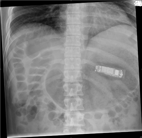
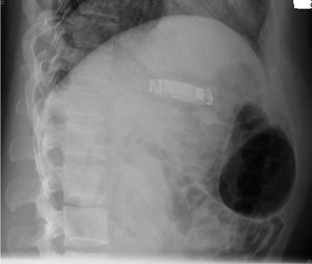
(such as abdominal tenderness, guarding or rigidity). An urgent erect chest and abdominal X-rays
were performed the same day in the Emergency department which confirmed the presence of the
mobile phone in the stomach (Figure 1 and 2). The stomach looked distended (seen on both the
antero-posterior and lateral views) but there was no free gas under the diaphragm on the erect chest
radiograph to suggest perforation.
The patient was kept overnight for observation, with no acute
concerns reported during his stay. He was reviewed the next morning
and his case was discussed with the upper GI consultant surgeon for
possible endoscopic retrieval of the phone. As there was no evidence
of peritonism or peritonitis, it was decided to discharge the patient
home and was booked on the next available therapeutic endoscopy
list. The phone dimensions were confirmed before the procedure by
looking up the phone details over the internet as well as the X-ray
radiograph. The patient was advised to take a soft diet and report
to the A&E department in the event of worsening symptoms while
waiting for the endoscopy.
The therapeutic upper GI endoscopy was performed five days
later. The procedure was performed under Xylocaine local oral spray
and heavy sedation (Fentanyl 100 micrograms; midazolam 2 mg). The
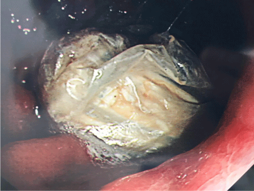
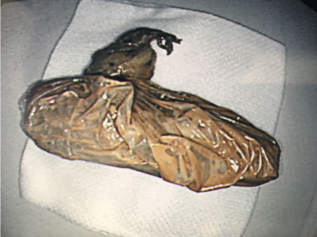
mobile phone was successfully retrieved endoscopically using a large endoscopic snare without any immediate or delayed complications.
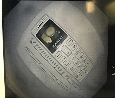
The plastic wraps were still intact containing the phone (Figure 3 and
4). The mobile phone retrieved was confirmed to be Zanco Mini with
0.66 inch LCD screen. The dimensions of the phone were: 71.8 mm x
23.5 mm x13.0 mm and weighed about 20 grams (Figure 5).
Although the procedure was uncomfortable for the patient,
re-scoping did not show any mucosal damage to the oesophagus
or gastro-oesophageal junction. The patient had a smooth postprocedure
recovery and was discharged home the same day with no
further follow-up.
Discussion
Intentional or accidental ingestion of foreign bodies is common
in the pediatric population and not unusual in adults. Most patients
are asymptomatic and the foreign bodies will pass through the gastrointestinal
tract without causing any significant harm, and these
patients will usually settle with reassurance and advice. However,
some patients will present with odynophagia, dysphagia, nausea,
saliva drooling, vomiting, chest and abdominal pain, and these
symptoms will depend on the type of the object ingested. Sharp
objects may cause bleeding, fistula formation and visceral perforation.
Even acute appendicitis has been reported as a result of foreign
body ingestion [5]. Large objects may also cause bowel obstruction
necessitating exploratory laparotomy. The common sites of foreign
body impaction are crico-pharyngeus, gastro-oesophageal junction,
pylorus and the ileo-caecal valve. These patients will need a thorough
history taking and clinical examination on presentation. Small objects
stuck at the crico-pharyngeal level may be removed in the A&E
department by an experienced clinician. However, other patients will
require radiological or endoscopic investigations. Plain X-rays of the neck, chest and/or abdomen is a useful early investigation to localize
the object. Further radiological follow-up may be required if the
foreign body is radio-opaque in order to follow the movement of the
object and to confirm its exit from the body. CT scan of the chest and
abdomen may be needed in the difficult or clinically significant cases.
A handheld metal detector has been shown to be safely and reliably
used in lieu of plain radiography to investigate children with a history
of metallic foreign body ingestion [6].
Mobile phone ingestion is unheard of in medical history. The
patient in our case report had wrapped the phone in two layers
of plastic bags before ingestion and therefore had no serious
consequences as the phone battery was still intact and not affected
by the stomach juices, however, the mere presence of the phone
could have caused gastric perforation due to its pressure effect. We
were comfortable to wait for 5 days to get the case done on an ideal
therapeutic list performed by an upper GI surgeon due to the fact that
the phone was wrapped in plastic covers and also, necessary surgical
expertise was available in case of gastric or oesophageal perforation.
Also, the size of the mobile phone in our case report was small but
we believe that any bigger size would have been extremely difficult
to be swallowed in the first place. We recommend that all such cases
should be investigated with early flexible endoscopy and assessed for
endoscopic retrieval. It is crucial that the procedure is carried out by
an experienced endoscopist with necessary interventional expertise
and arrangements made ready for surgical exploration in case of
failed endoscopic intervention. Our case report highlights a potential
problem in the modern era of growing technology and manufacture,
and opens a window for discussion on the best clinical management
of such cases. We anticipate that with popularity of the mobile phones
and the utter dependence on them, more such cases will be reported.
Figure 1
Figure 2
Figure 3
Figure 4
Figure 5
Learning Point
Clinicians need to be aware of the variety of objects that can be intentionally or accidentally swallowed, especially the gadgets of the modern world and the associated complications that these objects pose. Upper GI endoscopy is an important tool to investigate and retrieve ingested foreign bodies. It is important to get a good idea of the size and dimensions of the ingested object before retrieval is attempted in order to avoid visceral damage or perforation, and arrange surgical exploration if required.
Acknowledgement
Dr Khalid Mahmood (Radiology department) for his report and advice on X-rays.
References
- Blaho KE, Merigian KS, Winbery SL, Park LJ, Cockrell M. Foreign body ingestions in the emergency department: case reports and review of treatment. J Emerg Med. 1998; 16: 21-26.
- Ikenberry SO, Jue TL, Andersonetal MA. Management of ingested foreign bodies and food impactions. Gastrointest Endosc. 2011; 73: 1085-1091.
- Chih-Chien Yao, I-Ting Wu, Lung-Sheng Lu, Sheng-Chieh Lin, Chih- Ming Liang, Yuan-Hung Kuo, et al. Biomed Res Int. 2015.
- Sugawa C, Ono H, Taleb M, Lucas CE. Endoscopic management of foreign bodies in the upper gastrointestinal tract: a review. World J Gastrointest Endosc. 2014; 10: 475-481.
- Joo Heung Kim, Dae Sup Lee, Kwang Min Kim. Acute appendicitis caused by foreign body ingestion. Ann Surg Treat Res. 2015; 89: 158-161.
- Ramlakhan SL, Burke DP, Gilchrist J. Things that go beep: experience with an ED guideline for use of a handheld metal detector in the management of ingested non‐hazardous metallic foreign bodies. Emerg Med J. 2006; 23: 456-460.

