Case Report
Direct Tracheobronchial Suction: A Simple Technique to Relieve Post-Extubation Atelectasis in an Extremely Low Birth Weight Infant
Wan-Jung Tsai1, Mei-Jy Jeng1,2,3*, Pei-Chen Tsao1,2,3, Yu-Sheng Lee1,2,3 and Wen-Jue Soong1,2,3
1Department of Pediatrics, Taipei Veterans General Hospital, Taiwan
2Institute of Emergency and Critical Care Medicine, National Yang-Ming University, Taiwan
3Department of Pediatrics, National Yang-Ming University, Taiwan
*Corresponding author: Mei-Jy Jeng, Institute of Emergency and Critical Care Medicine, School of Medicine, National Yang-Ming University, Taipei 112, Taiwan
Published: 11 Oct, 2016
Cite this article as: Tsai W-J, Jeng M-J, Tsao P-C, Lee Y-S,
Soong W-J. Direct Tracheobronchial
Suction: A Simple Technique to Relieve
Post-Extubation Atelectasis in an
Extremely Low Birth Weight Infant. Ann
Clin Case Rep. 2016; 1: 1155.
Abstract
Post-extubation atelectasis is a common pulmonary complication of critical neonates who have
received long-term, high-setting of mechanical ventilator, resulting in re-intubation, increased
morbidity and lengthened hospitalizations. A rapid and effective management to open the lungs
is important to prevent reintubation. We reported an extremely preterm infant with massive postextubated
atelectasis of bilateral lungs, and he was successfully treated by using the simple technique
of direct tracheobroncheal suction (DTBS) to quickly open his lungs.
A 7-day-old preterm infant with a birth weight of 732 g got massive atelectatic lungs at 24 hours
after extubation. DTBS was performed by directly inserting a 6.0 Fr. suction tube into his trachea via
the assist of laryngoscope. The infant’s head was turned to one side while passing the suction tube
into the bronchus of the opposite side. This suction procedure was repeated until nothing could be
suctioned out, and DTBS was repeated every 8 hours in the following 24 hours after extubation. Nasal
prong continuous positive airway pressure was kept, and the heart rates and oxygen saturation were
continuously monitored during DTBS. Sticky secretions were observed at the laryngeal site and
suctioned out from the tracheobronchial airways. The follow-up chest x-ray film revealed opened
lungs 24 hours later. Therefore, DTBS is effective and safe to relief obstructed airways and reopen
the lungs of the reported extremely premature infant.
Keywords: Post-extubation atelectasis; Direct tracheobronchial suction
Introduction
Post-extubation atelectasis is a common pulmonary disorder that lengthens the hospitalization
time of neonates in intensive care units who have received long-term, high-setting of mechanical
ventilator, resulting in increased morbidity among these infants. The incidence of post-extubation
atelectasis has been reported in 16.7 to 50% of intubated neonates, especially in those premature
infants group [1,2].
The current conventional prevention strategies and management of post-extubation atelectasis
including mucolytic medication, chest physiotherapy, but poor response and re-intubation rate
in neonates, especially in premature infants are still high which has been reported ranged from
18 to 34% [1-7]. Some reports have been documented and suggested the use of bronchoscope
to check and relieve that intractable atelectasis. However, technical limitation and instrument
limitation of bronchoscope may made difficulties in many institutes to perform those invasive
techniques, especially performed in neonates or premature infants [6]. A simple technique of direct
tracheobronchial suction (DTBS) had been reported for relieving post-extubated atelectatic lungs
in premature infants [6].
DTBS could achieve direct clearance of trachea and bronchus by multiple insertions, natural
curvature of suction tube tip and head positioning without the need of re-intubation. The
effective airway secretion removal could predict good results of resolving the massive atelectasis
and preventing the possibility of re-intubation. There are some immediate findings such as
respiratory distress improved with increased audible air entry on the affected lung; decreased chest
retractions; significant fall in respiratory rate and heart rate; arterial blood gases analysis showed significant improvement of pH, partial pressure of carbon dioxide
and oxygenation ratio and partial or nearly complete resolution of
the atelectasis by chest radiograph check-up. Besides, the aspirated
secretion in suction tube could also send for laboratory study if
needed [6].
Here, we present an extremely pretrem infant who suffered
from massive lung atelectasis after extubation and got significant
improvement 24 hours after being performed with DTBS.
Case Presentation
The case is a male infant delivered at gestational age of 28
weeks by cesarean section from a 33-year-old woman due to severe
preeclampsia. His birth weight was 732 gm, and the Apgar scores
were 3, 6 and 8 at 1, 5, and 10 min after delivery. After birth, he got
severe respiratory distress with subcostal retraction and cyanosis, so
oral endotracheal tube was inserted and he was ventilated with highfrequency
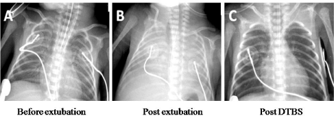
oscillatory ventilation. The chest x-ray film revealed grade
III respiratory distress syndrome (RDS) (Figure 1A). The pulmonary
conditions got improved gradually, and the chest x-ray film showed
a good improvement. Extubation was done on the 7th day of life.
Nasal prong continuous positive airway pressure (NCPAP) was
applied immediately after extubation. Noninvasive positive pressure
ventilation (NIPPV) (rate =20 breaths/min, peak inspiratory pressure
= 18cm H2O, positive end expiratory pressure = 6cm H2O, and FiO2
= 0.3) were applied. Under these settings, these was no significant
hypercapnia or hypoxia observed during the first 12 hours after
extubation. However, relative fluctuation and occasional dropping
of the oxygen saturation to lower than 90% started to be observed
at night. The FiO2 needed to be increased to 0.4 for maintaining
oxygen saturation higher than 90%. Mild tachypnea and subcostal
retraction of the infant were also observed. The blood gas also showed
compensatory respiratory acidosis with a tendency of increasing
PaCO2. Chest x-ray film was rechecked at approximately 24 hours
after extubation, and it showed white-out appearance with decreased
lung volume at bilateral lung fields. Massive post-extubation
atelectasis was considered (Figure 1B).
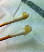
By the use of a laryngoscope, the oral cavity and larynx were
checked firstly. Much sticky secretion was observed and removed by
using cotton swabs (Figure 2). Then, DTBS was performed under the
continuous support of NCPAP. Three minutes before the procedure,
the FiO2 was increased 20% higher than before for pre-oxygenation.
While performing DTBS, we inserted a 6.0 Fr. suction tube into
his trachea directly via the assist of laryngoscope. The infant’s head
was put on neutral position while inserted the tube into the vocal
cord. While reaching the vocal cord, the infant’s head was turned to right side firstly and the suction tube was passed deeper into left
bronchus until a resistance was felt. Then, the suction tube was slowly
withdrawn out with a suction pressure of 80 mmHg. This suction
procedure would be repeated again until nothing could be suctioned
out. After that, we suctioned the right bronchus with head turned to
the left side until the airway is clear. Between each two suctions, we
kept the mouth closed and waited for the oxygenation turning back
to be higher than 95%. During the procedures, transient oxygen
desaturation and brief bradycardia were observed occasionally, and
they returned back quickly. Topical epinephrine was applied over
larynx after whole procedure for laryngeal swelling. The DTBS was
performed again in an interval of 8 hours.
The follow-up chest x-ray film revealed opened lungs 24 hours
later (Figure 1C). After that, DTBS was performed twice a day for two
more days. His respiratory condition got improved and the settings of
NIPPV were able to be weaned gradually.
When the patient was 28 days old, the baby still required NCPAP
with NIPPV. At postmenstral age of 36 weeks, he still required
the support of high-flow nasal cannula oxygen to maintain a nasal
continuous positive airway pressure with 3cm H2O PEEP and
oxygen (FiO2 = 0.25), so he was diagnosed as moderate to severe
bronchopulmonary dysplasia (BPD) of prematurity. With a good
care, neither NCPAP nor oxygen was required 3 weeks later, and he
was discharged at postmenstral age of 40 weeks with body weight
reaching 2100g. The patient was under regular out-patient follow-up
with generally well-being.
Figure 1
Figure 1
Chest plain film of the presented premature infant. (A) Before extubation. (B) 24 hours after extubation. Massive pulmonary atelectasis at bilateral lung
fields was shown. (C) 24 hours after performing direct tracheobroncheal suction. It showed well-expanded lungs bilaterally.
Figure 2
Figure 2
Thick and glutinous secretion removed by cotton swabs from the
laryngotracheal orifice of the presented premature infant.
Discussion
Atelectasis is known as the loss of lung volume due to the collapse of lung tissue, and the most type in adult and children is obstructive
type. The critical illness neonates, especially premature infant in
intensive care unit usually require intubation with mechanical
ventilation support and frequent secretion suction [1,2]. There are
some researches had documented the recent ventilator strategy with
high frequency oscillatory ventilator compared with conventional
ventilation in premature infants, and better successful extubation
rate had reported with premature infants who use high frequency
oscillatory ventilator and the age at successful extubation was also
significantly lower compared with those who use conventional
ventilator [8-11]. But, there are still many risk factors played
important role in atelectasis process causing alveolar collapse, such
as neonates are sensitive to the obstructing effects of accumulating
airway secretions, most likely because of small airway size and a
less effective cough secondary to muscle weakness; endotracheal
tubes complicate this problem by impairing mucocillary clearance,
inhibiting an effective cough, causing airway mucosa damage and
granulation tissue formation; neonates have a decreased number
of pores of Kohn which limits collateral ventilation [6,7]. The high
incidence of post-extubation atelectasis has been reported in 16.7
to 50% of intubated neonates, especially in those premature infants
group, result from above risk factors [1,2].
Due to the above risk factors and the most cause of airway
secretions, the goals of prevention and management of postextubation
atelectasis is remove airway secretion as soon as possible
to keep patent airway and avoid subsequent pulmonary dysfunction.
Traditionally, the treatment strategies for post-extubation atelectasis
of intubated neonates were mucolytic medication and intensive
chest physiotherapy. However, tiny neonates and premature infants
often could not stand for fierce percussion movement, and would
sometimes lead to brain damage or bleeding tendency. Generally, if
the massive post-extubation atelectasis could not be resolved by chest
physiotherapy, re-intubation was necessary for following respiratory
support and secretion clearance and got temporary improvement.
But, the subsequent trauma processes caused by endotracheal tubes
and suction tubes would lead to further more atelectasis episode after
extubation and add up to a vicious cycle of pulmonary destruction
which affected pulmonary development of fragile neonates and
premature infants [6].
Bronchoscopy with or without endotracheal tube is a useful
technique to achieve diagnosis and treatment for atelectasis.
However, premature infants may not stand for the procedure under
general anesthesia, and their airway may too small to receive a
standard pediatric bronchoscope. Therefore, direct tracheobronchial
suction seen to be the better choice to achieve direct tracheobronchial
clearance without the need of re-intubation and the use of
bronchoscope. Under the bedside monitors for basic vital sign and
saturation, and with a laryngoscope and a suction tube, we could
precisely perform direct clearance of trachea and bronchus by
multiple insertions, natural curvature of suction tube tip and head
positioning. The effective airway secretion removal could predict
good results of resolving the massive atelectasis and preventing the
possibility of re-intubation. There are some immediate findings such
as respiratory distress improved with increased audible air entry
on the affected lung; decreased chest retractions; significant fall in
respiratory rate and heart rate; arterial blood gases analysis showed
significant improvement of pH, partial pressure of carbon dioxide and oxygenation ratio and partial or nearly complete resolution of
the atelectasis by chest radiograph check-up. Besides, the aspirated
secretion in suction tube could also send for laboratory study if
needed.
After the procedure, the premature infants must be intensive
monitor for any acute complication such as hypoxia, pneumothorax,
pulmonary bleeding. Because even flexible suction tube which diameter
is smaller than bronchoscope could also damage airway mucosa and
moreover perforations. Besides, intensive chest physiotherapy is still
recommended even receive direct tracheobronchial suction for better
airway clearance [6].
In conclusion, extremely premature infants have high risk to
develop atelectasis after extubation. DTBS is a simple, effective and
less invasive procedure to resolve post-extubation atelectasis and
prevent re-intubation in tiny infants. Therefore, DTBS may be tried
on premature infants with post-extubation atelectasis.
Acknowledgment
Supported in part by grants from Taipei Veterans General Hospital(V105C-182) and Ministry of Science and Technology (MOST 105-2314-B-010-044), Taipei, Taiwan.
References
- Lúcia Cândida Soares de Paula, Fernanda Corsante Siqueira, Regina Célia Turola Passos Juliani, Werther Brunow de Carvalho, Maria Esther Jurfest Rivero Ceccon, Uenis Tannuri. Post-extubation atelectasis in newborns with surgical diseases: a report of two cases involving the use of a high-flow nasal cannula. Revista Brasileira de terapia intensive. 2014; 26: 317-320.
- Lee CY, Su BH, Lin TW, Lin HC, Li TC, Wang NP. Risk factors of extubation failure in extremely low birth weight infants: a five year retrospective analysis. Acta paediatrica Taiwanica. 2002; 43: 319-325.
- Principi T, Fraser DD, Morrison GC, Farsi SA, Carrelas JF, Maurice EA, et al. Complications of mechanical ventilation in the pediatric population. Pediatr Pulmonol. 2011; 46: 452-457.
- Mayordomo-Colunga J, Medina A, Rey C, Concha A, Menéndez S, Los Arcos M, et al. Non invasive ventilation after extubation in paediatric patients: a preliminary study. BMC Pediatr. 2010; 10: 29.
- Schechter MS. Airway clearance applications in infants and children. Respir Care. 2007; 52: 1382-1390.
- Soong WJ, Jeng MJ, Hwang B. Direct tracheobronchial suction for massive post-extubation atelectasis in premature infants. Zhonghua Minguo xiao er ke yi xue hui za zhi. 1996; 37: 266-271.
- Al-Alaiyan S, Dyer D, Khan B. Chest physiotherapy and post-extubation atelectasis in infants. Pediatr Pulmonol. 1996; 21: 227-230.
- Zivanovic S, Peacock J, Alcazar-Paris M, Lo JW, Lunt A, Marlow N, Calvert S. Late outcomes of a randomized trial of high-frequency oscillation in neonates. N Engl J Med. 2014; 370: 1121-1130.
- Dargaville PA, Tingay DG. Lung protective ventilation in extremely preterm infants. J Paediatr Child Health. 2012; 48: 740-746.
- Johnson AH, Peacock JL, Greenough A, Marlow N, Limb ES, Marston L, et al. High-frequency oscillatory ventilation for the prevention of chronic lung disease of prematurity. N Engl J Med. 2002; 347: 633-642.
- Courtney SE, Durand DJ, Asselin JM, Hudak ML, Aschner JL, Shoemaker CT et al. High-frequency oscillatory ventilation versus conventional mechanical ventilation for very-low-birth-weight infants. N Engl J Med. 2002; 347: 643-652.


