Clinical Image
Initial Presentation of Tuberous Sclerosis Complex in an Adult Woman with Hemorrhage from Bilateral, Giant, Multi-Focal Renal Angiomyolipomas
Carrie Stewart1*, Ashley Richman2, Jacob Scheibal3 and Jonathan Silberstein1
1Department of Urology, Tulane University, USA
2Department of Urology, The University of Queensland School of Medicine, USA
3Department of Radiology, Ochsner Medical Center, USA
*Corresponding author: Carrie Stewart, Department of Urology, Tulane University, 1430 Tulane Avenue, SL-42, New Orleans, LA 70112, USA
Published: 28 Sep, 2016
Cite this article as: Stewart C, Richman A, Scheibal J, Silberstein J. Initial Presentation of Tuberous Sclerosis Complex in an Adult Woman with Hemorrhage from Bilateral, Giant, Multi-Focal Renal Angiomyolipomas. Ann Clin Case Rep. 2016; 1: 1147.
Abstract
Renal angiomyolipomas (AML’s) are benign tumors containing smooth muscle, adipose, and aberrant vasculature and can cause hypertension, renal failure and life threatening hemorrhage. In Tuberous sclerosis complex (TSC), AMLs are often bilateral and can be too numerous to count, up to 89% and 76% respectively. The disease is autosomal dominant and affects multiple anatomic systems. No single feature is diagnostic for TSC, making complete evaluation for major and minor signs requisite. We report a case of a hemorrhagic AML in a woman with giant, bilateral, multifocal AML and hypovolemic shock requiring emergent endovascular treatment of unilateral AML hemorrhage.
Keywords: Angiomyolipoma; Sirolimus; mTOR; Giant angiomyolipoma
Clinical Image
During workup for diminished appetite and weight loss, a 53 year-old woman was found to
have bilateral giant AML’s. Severe anemia was discovered on labs drawn for symptoms of dizziness
and fatigue; she was referred for emergency treatment. Because her eGFR was 20, she underwent a
non-contrasted CT. This demonstrated mixed attenuation within an 11 cm superior, left AML and
lymphangiomyomatosis, a second major and confirmatory sign of TSC (Figure 1).
After 3 units of packed red cells and fresh frozen plasma, her hematocrit did not respond.
Angiogram demonstrated a large pseudoaneurysm with an arteriovenous fistula arising from the
left renal artery, in the superior lateral AML. The pseudoaneurysm was embolized using Ruby® coils (Penumbra Inc., Alameda, CA). Subsequently, the artery feeding the
AML of the left upper pole was embolized with alcohol and lipidol.
Post embolization imaging demonstrates the successfully treated
pseudoaneurysm with appropriate blood flow to the left kidney
(Figure 2).
Her creatinine, hemoglobin and hematocrit stabilized after
embolization. Repeat CT demonstrated hematoma stability, and
no further transfusions were required. Following discharge she was
scheduled to see oncology for Sirolimus, an mTOR inhibitor, which
is FDA approved to treat AMLs and may result in regression [1-4].
Figure 1
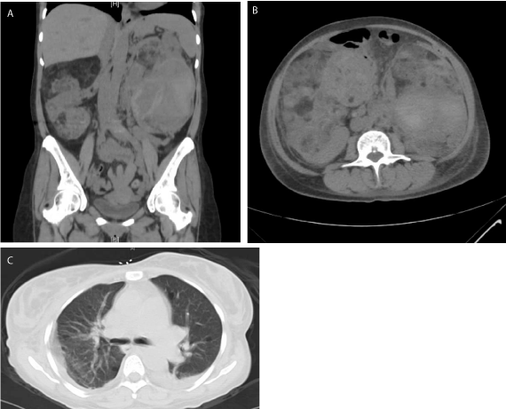
Figure 1
(a) Non-contrast coronal (b) axial CT showing bilateral angiomyolipomas essentially replacing her
kidneys bilaterally with a suspected left retroperitoneal hematoma (arrow) (c) Scattered pulmonary cyst (arrows)
suggestive of lymphagiomyomatosis, also seen in tuberous sclerosis.
Figure 2
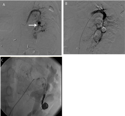
Figure 2
(a) Angiography of the left kidney with pseudoaneurysm (arrow)
arising from the superior pole of the left kidney with extravasation of
contrast inferiorly. (b) Post-interventional angiography demonstrating coiled
pseudoaneurysm with resolution of contrast extravasation. (c) Angiography
of the left renal artery after embolization with alcohol and lipiodol showing
decreased perfusion to left upper pole AML.
References
- Peng, Zhu-Feng, Lu Yang, Ting-Ting Wang, Ping Han, Zhen-Hua Liu. Efficacy and Safety of Sirolimus for Renal Angiomyolipoma in Patients with Tuberous Sclerosis Complex or Sporadic Lymphangioleiomyomatosis: A Systematic Review. J Urol. 2014; 192: 1424-1430.
- Bissler John J, Francis X Mccormack, Lisa R Young, Jean M Elwing, Gail Chuck, Jennifer M Leonard, et al. Sirolimus for Angiomyolipoma in Tuberous Sclerosis Complex or Lymphangioleiomyomatosis. N Engl J Med. 2008; 358: 140-151.
- Casper Keith A, Lane F Donnelly, Bin Chen, John J Bissler. Tuberous Sclerosis Complex: Renal Imaging Findings1. Radiology. 2002; 225: 451- 456.
- Crino Peter B, Katherine L Nathanson, Elizabeth Petri Henske. The Tuberous Sclerosis Complex. N Engl J Med. 2006; 355: 1345-1356.