Clinical Image
Idiopathic Median Canaliform Dystrophy of Heller Involving Both Thumb Nails
Pragya Ashok Nair*
Department of Skin & VD, Pramukhswami Medical College, India
*Corresponding author: Pragya Ashok Nair, Department of Skin & VD, Pramukhswami Medical College, Anand Sojitra Road, Karamsad, Anand, Gujarat 388325, India
Published: 19 Sep, 2016
Cite this article as: Nair PA. Idiopathic Median Canaliform
Dystrophy of Heller Involving Both
Thumb Nails. Ann Clin Case Rep. 2016;
1: 1140.
Clinical Image
A 16 year old boy presented to us with asymptomatic exfoliation of skin over both lateral
and proximal thumb folds. No history of biting of nails was elicited as during stress. No history
of contact with irritants or allergens was present. No specific family history was elicited. No skin
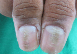
lesions present over any part of body. On examination, both the thumb nails showed a median
longitudinal groove extending from proximal nail fold to the distal nail edge with transverse furrows
arising on either side (Figure 1). Lunula was enlarged in size. Exfoliation was present over both
lateral and proximal nail fold. Rest other finger and toe nails were normal. Systemic examination
showed no abnormality. Potassium mount prepared from the scraping of nails were negative for
fungal elements. Biopsy was not done as patient refused for it. Diagnosis of median nail dystrophy
was made on clinical basis and patient was put on 0.1% tacrolimus ointment topically at night, but
patient didn’t return in follow up.
Median canaliform dystrophy (MCD) of Heller also known as solenonychina, dystrophia
unguis mediana canaliformis, and nevus striatus unguis is a rare entity characterized by a midline
or a paramedian ridge or split and canal formation in nail plate of one or both the thumb nails
[1]. It rarely involves toe nails and other finger nails. The majority of cases of median canaliform
dystrophy are idiopathic. Other causes includes traumatic injury to the base of nails, use of
oral retinoids, Subungual skin tumors, such as glomus, myxoid, and other tumors resulting in
longitudinal grooving and lifting of the nail plate from the bed [2]. Sweeney et al. [3] reported a
familial clustering of cases of median nail dystrophy [4].
The first case was recorded by Heller in 1928 [1]. There is no sex predilection. Mean age of
occurence is 25.72 years. The condition is diagnosed based on its clinical features. It results from a
temporary defect in the nail matrix, following dyskeratinization or focal infection, or due to selfinflicted
trauma to the nail plate, nail matrix or nail bed [3]. It presents with small cracks or fissures
that extend laterally from the central canal or split towards the nail edge giving the appearance of an
inverted fir tree or Christmas tree, usually symmetrically affecting the thumb nails mainly [4]. There is absence of keratinocytes adhesions with in nail matrix with dyskeratosis which is responsible for
formation of longitudinal grove with splitting of nail plate due to weaker tensile strength.
The habit tic deformity is the closest differential which produces transverse ridges along the
central nail plate depression instead of a longitudinal groove with lateral projections as seen in MCD.
The treatment of MCD depends on the etiology. As in majority of cases the cause is unknown and it may revert back to normal after many months to years as was seen in
our case where we could not find the cause. Injectable triamcinalone
acetonide, topical 0.1% tacrolimus, and tazarotene 0.05% cream are
other options with variable results [5]. Psychiatric opinion should be
taken when associated with the depressive, obsessive-compulsive, or
impulse-control disorder [6]. Any underlying tumour if any needs to
be removed.
A case of median nail dystrophy is also reported by Madke B et
al. [7] as our case who was a 16 year old young adolescent boy, which
is the age when stressful event in life can lead to depression and thus
patient inflicts trauma manipulating the cuticular portion of nail fold.
Such history could not be elicited in our patient even after taking him
in confidence, so we labeled it as idiopathic case of MCD.
Figure 1
Figure 1
Median longitudinal groove with fissures arising on either side giving a fir tree appearance with
enlarged lunula and exfoliation of skin involving both thumb nails.
References
- Beck M, Wilkinson S. Disorders of nails: Medican canaliform dystrophy. In: Burns T, Breathnach S, Cox N, Griffiths C, editors. Rook’s Textbook of Dermatology. 7th ed. Oxford: Blackwell Science; 2004: 54-55.
- Verma SB. Glomus tumor-induced longitudinal splitting of nail mimicking median canaliform dystrophy. Indian J Dermatol Venereol Leprol. 2008; 74: 257-259.
- Sweeney SA, Cohen PR, Schulze KE, Nelson BR. Familial median canaliform nail dystrophy. Cutis. 2005; 75: 161-165.
- Wu CY, Chen GS, Lin HL. Median canaliform dystrophy of Heller with associated swan neck deformity. J Eur Acad Dermatol Venereol. 2009; 23: 1102-1103.
- Kim BY, Jin SP, Won CH, Cho S. Treatment of median canaliform nail dystrophy with topical 0.1% tacrolimus ointment. J Dermatol. 2010; 37: 573-574.
- Kota R, Pilani A, Nair PA. Median Nail Dystrophy Involving the Thumb Nail. Indian J Dermatol. 2016; 61: 120.
- Madke B, Gadkari R, Nayak C. Median canaliform dystrophy of Heller. Indian Dermatol Online J. 2012; 3: 224-225.

