Case Report
Mineral Trioxide Aggregate as an Apical Plug in Endodontic Management of Non-Vital Permanent Teeth with Open Apex: A Case Report
Camargo Carlos H*, Khoury Rayana D, Valera Marcia C, Carvalho Cláudio A and Torres
Carlos R
Department of Restorative Dentistry, State University of São Paulo, Brazil
*Corresponding author: Carlos H R Camargo, Endodontics Division, Universidade “Júlio de Mesquita Filho” – UNESP, Av. Eng. Francisco José Longo, nº 777, Jardim São Dimas - 12245-000, São José dos Campos, SP, Brazil
Published: 01 Nov, 2016
Cite this article as: Camargo Carlos H, Khoury Rayana
D, Valera Marcia C, Carvalho Cláudio
A, Torres Carlos R. Mineral Trioxide
Aggregate as an Apical Plug in
Endodontic Management of Non-Vital
Permanent Teeth with Open Apex: A
Case Report. Ann Clin Case Rep. 2016;
1: 1134.
Abstract
Apexification is a widely used technique to manage immature permanent teeth and can be performed either using calcium hydroxide (Ca(OH)2) or mineral trioxide aggregate (MTA). This paper illustrates a case report of a unsuccessful apexification with Ca (OH)2 of amaxillary incisor that was then treated by an apical barrier made of MTA.
Keywords: Mineral trioxide aggregate; Immature teeth; Apexification
Introduction
In Endodontics, one of the most significant difficulties is the management of immature necrotic
teeth. The apical closure is essential to endodontic therapy because it properly seals the root canals
system as well as maintains the filling material restricted to the root canals. Traditionally, such
conditions is treated using calcium hydroxide in order to induce apexification and by that establish
a natural constriction of the apical portion [1,2]. However, the need of multiple appointments
illustrates a relevant disadvantage regarding the use of calcium hydroxide. Besides that, the period
time required for treatment achievement using Ca(OH)2 may range from 5 to 20 months [3-
5] which is considered a long-term therapy and it can be associated to many risks such as root
weakening and tooth fracture [6]. To overcome prolonged treatment time related to the technique
mentioned above, mineral trioxide aggregate (MTA) has been reported to be an effective material
for root end filling due to its excellent sealing ability and biocompatibility [7,8]. Further more, MTA
also promotes the hard tissue formation, which enhances its potential to prevent leakage of bacteria
and their by-products [9].
Considering these properties and taking into account the lack of patient motivation to attend
to multiple visits, the MTA apical plug placement has been suggested. In addition to its several
favorable characteristics, the insertion of MTA in the apical end of the root canal enables the
treatment to be achieved more quickly since the root canal can be immediately filled [2].
This case report presents unsuccessful apexification with Ca(OH)2 more than 20 months and
then a successful MTA apical plug placement in an maxillary central incisor with pulp necrosis and
open apex.
Case Presentation
A 13-year-old female presenting good general health was referred to continue root canal
treatment that had been started 3 years earlier due to a dental trauma suffered in the left maxillary
central incisor. Clinical examination revealed crown fracture, coronary darkening and absence of
abnormal mobility (Figure 1A). The tooth was unresponsive to cold test (Endo-Ice, The Hygenic
Corp.,Akron, OH, USA). Radiographic examination (Figure 1B) revealed an immature tooth 21,
with wide canal, open apex and extensive radiolucent lesion around the periapex. The apexification
procedure was explained to the patient's parents and they consented to the treatment.
After anesthesia and rubber dam placement, endodontic access was performed following
conventional guidelines. The coronal and middle thirds were then irrigated using saline solution
in order to remove remnant intracanal medication placed 3 years before. The working length was
determined, and the apical third dentin walls were cleaned up to a K-file # 80 (Dentsply Maillefer,
Ballaigues,Switzerland). The canal was irrigated with copious amounts 1% sodium hypochlorite irrigation with negative apical pressure with NaviTip tips and dried
by capillary tips (Ultradent, South Jordan, UT). A calcium hydroxide
paste with aqueous vehicle was inserted into the apical portion of
canal with a spiral lentuloas intracanal medication. The access cavity
was sealed with glass ionomer. Periodic changes (up two months) of
the root canal dressing material were made for more than 20 months
(using calcium hydroxide powder mixed to propylene glycol vehicle).
After this period, on a follow-up visit, it was observed the remission
of radiolucent image, which meant the initial healing of the periapical
lesion. On the other hand, calcium hydroxide apexification was unable
to adequately form an apical root canal barrier. Due to the extent of
the root canal width and the lack of an apical stop barrier, a novel
endodontic treatment using mineral trioxide aggregate (MTA) as an
apical plug was preconized. The intracanal medication was removed
by repeated irrigation with saline solution and the root canal was
dried with absorbent paper points (Dentsply, Petrópoilis-RJ, Brazil).
MTA (Angelus, Londrina-PR, Brazil) was prepared according to the
manufacturer’s instructions and placed in the apical portion of root
canal (3-4 mm) using the MTA carrier (1.6 mm, Dovgan Tip, USA),
creating an apical barrier. Periapical radiograph was taken to confirm
the correct position and size of the MTA plug (Figure 2A). Remaining
root canal space was obturated by the (lateral condensation technique
using gutta-percha modeled main point (Dentsply-Maillefer,
Ballaigues, Switzerland) (Figure 2B), with AHPlus (Dentsply) as a
endodontic sealer. Finally, obturation was partially removed (Figure
2C) and the tooth was restored with fiber posts and composite resin
core (F250 3M-ESPE, St. Paul, MN, USA--------) (Figure 2D) 5.
One week after the patience receives dental bleaching and esthetic
restoration (Figure 3A). At both 22 months and four-year follow-up,
patient was clinically asymptomatic and the periapical lesion had
disappeared (Figure 3B).
Figure 1
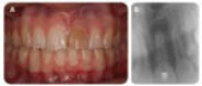
Figure 1
(a) Extra-oral pre-operative image of child. (b) A preoperative
periapical radiograph.
Figure 2
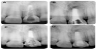
Figure 2
(a) Radiograph confirmation of the correct position and size of the
MTA plug. (b)Obturation of the root canal space by thelateral condensation
technique using gutta-percha modeled main point. (c) Obturation partially
removed to place the fiber posts and composite resin core. (d) Fiber posts
and composite resin core placed.
Figure 3
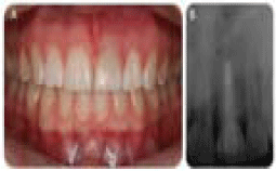
Figure 3
(a) Extra-oral post-operative image of child. (b) At the 4-year follow
up the periapicallesion had disappeared.
Discussion
The prognosis of a dental injury is uncertain, especially when
it involves immature teeth, because it may become non-vital and infected. The loss of its blood supply interrupts the continued
maturation and the apical closure of the root canal [10]. In Endodontic
field the treatment of injured permanent immature teeth presents an
exceptional challenge faced by endodontists due to the thin and fragile
dentine walls. This paper illustrates unsuccessful apexification closure
with Ca(OH)2 along 18 months of an immature maxillary incisor that
was then successfully treated with MTA as root end filling material.
For many decades apexification using calcium hydroxide has been
the treatment of choice aiming the formation of hard tissue barrier
and healing in non-vital immature permanent teeth [11]. However,
the long treatment time associated to this technique constitutes a
relevant drawback once it depends on the patient’s cooperation to
attend several follow-up appointments [6,12]. Another issues related
to the use of calcium hydroxide for a long period is the risk of root
weakening along with tooth fracture [6,13] and coronal microleakage
during treatment [14]. Additionally, the formed barrier achieved
during apexification, usually appears to be calcified, though it is
actually porous and may contains small amounts of softtissue [15].
On this regard, mineral trioxide aggregate (MTA) apical plug has
been proposed as an effectivetechnique for apical sealing of necrotic
immature teeth and has shown excellent results [16]. This material
is provided of excellent biocompatibility, antibacterial properties,
decreased apical leakage, better marginal adaptation, short setting
time (approximately 4 hours) and also has the capability to induct
hard tissue formation [17,18]. Furthermore, both the patient and
dentist benefit from the use of MTA since the whole treatment time
is greatly reduced [12].
Besides the apexification with calcium hydroxide and MTA apical
plug placement techniques, pulp revascularization is also among the
treatment options for necrotic immature teeth [19-21]. It involves
disinfecting the root canal system, providing a matrix of blood
clot into which cells could grow, and sealing of the coronal access.
Although, the decision for apexification instead of revascularization
was made primarily because the diameter of the open apex was not
more than 2 mm, which may be difficult to induce bleeding. All
cases of unfavorable revascularization outcome apparentlywere
related to a failure to induce any bleeding into the canal [22,23]. The
difficulty to achieve an impeccable control of bacterial infection also
seems to be highly relevant to the complexity and unpredictability
of the outcome of this procedure. Long-termclinical results are
as yet not available. Moreover, it is likely that the entire canalwill
be calcified, compromising esthetics and potentially increasethe
difficulty in future endodontic procedures, if required [24]. Thus,
now many endodontists are recommending revascularization just in cases where the root formation is in the very beginning, divergent
apex, these situations normally lead to tooth loss in the past due to
do not resist to occlusion forces. In the present case, postand core
were the final restorative treatment plan with bleaching, therefore
so, revascularization did not seem the right best treatment option
because the vital tissue in apical two thirds ofthe canal cannot be
violated for post placement.
For this case, MTA plug was an effective method for apical
sealing enabling posterior obturation. The apical plug placement was
performed in one single appointment, but the previous placement
of calcium hydroxide had significant improvement of the lesion
remission. It is worth mentioning that, iIn MTA plug technique,
a temporary calcium hydroxide medication should precede the
application of MTA to disinfect and restrict bacterial infection in the
root canals [13,25]. Otherwise, the Ca(OH)2 intracanal medication
can be maintained for only two weeks, differently from our case,
which the first option treatment was the apexification with calcium
hydroxide.
Additionally, there are some recent released materials like the
new MTA (Angelus, Londrina, Brazil) and Biodentine (Septodont,
Saint Maur des Fosses, France) that are reported to do not cause
discoloration, which is even better regarding its use as anapical plug.
However, new clinical studies need to be performed in order to
confirm better protocols.
Conclusion
Dental injuries in immature permanent teeth often result in endodontic complications. Apexification technique using calcium hydroxide is associated with certain flaws, such as long treatment time, the possibility of tooth fracture and incomplete calcification. The use of an apical plug employing mineral trioxide aggregate (MTA) is an alternative treatment option with the advantage of a shorter chairside treatment and immediate tooth reinforcement by fiber posts.
References
- AL Frank. Therapy for the divergent pulpless tooth by continued apical formation. J Am Dent Assoc. 1966; 72: 87-93.
- DR Morse, J O'Larnic, C Yesilsoy. Apexification: review of the literature. Quintessence Int. 1990; 21: 589-598.
- SJ Lee, M Monsef, M Torabinejad. Sealing ability of a mineral trioxide aggregate for repair of lateral root perforations. J Endod. 1993; 19: 541-544.
- DJ Kleier, ES Barr. A study of endodontically apexified teeth. Endod Dent Traumatol. 1991; 7: 112-117.
- A Dominguez Reyes, L Munoz Munoz, T Aznar Martin. Study of calcium hydroxide apexification in 26 young permanent incisors. Dent Traumatol. 2005; 21: 141-145.
- JO Andreasen, B Farik, EC Munksgaard. Long-term calcium hydroxide as a root canal dressing may increase risk of root fracture. Dent Traumatol. 2002; 18: 134-137.
- M Torabinejad, M Parirokh. Mineral trioxide aggregate: a comprehensive literature review--part II: leakage and biocompatibility investigations. J Endod. 2010; 36: 190-202.
- V Zand, M Lotfi, A Aghbali, M Mesgariabbasi, M Janani, H Mokhtari, et al. Tissue Reaction and Biocompatibility of Implanted Mineral Trioxide Aggregate with Silver Nanoparticles in a Rat Model. Iran Endod J. 2016; 11: 13-16.
- M Torabinejad, TF Watson, TR Pitt Ford. Sealing ability of a mineral trioxide aggregate when used as a root end filling material. J Endod. 1993; 19: 591-595.
- M Cvek. Prognosis of luxated non-vital maxillary incisors treated with calcium hydroxide and filled with gutta-percha. A retrospective clinical study. Endod Dent Traumatol. 1992; 8: 45-55.
- EC Sheehy, GJ Roberts. Use of calcium hydroxide for apical barrier formation and healing in non-vital immature permanent teeth: a review. Br Dent J. 1997; 183: 241-246.
- S Simon, F Rilliard, A Berdal, P Machtou. The use of mineral trioxide aggregate in one-visit apexification treatment: a prospective study. Int Endod J. 2007; 40: 186-197.
- JO Andreasen, EC Munksgaard, LK Bakland. Comparison of fracture resistance in root canals of immature sheep teeth after filling with calcium hydroxide or MTA. Dent Traumatol. 2006; 22: 154-156.
- WP Saunders, EM Saunders. Coronal leakage as a cause of failure in rootcanal therapy: a review. Endod Dent Traumatol. 1994; 10: 105-108.
- WH Binnie, AH Rowe. A histological study of the periapical tissues of incompletely formed pulpless teeth filled with calcium hydroxide. J Dent Res. 1973; 52: 1110-1116.
- DT Holden, SA Schwartz, TC Kirkpatrick, WG Schindler. Clinical outcomes of artificial root-end barriers with mineral trioxide aggregate in teeth with immature apices. J Endod. 2008; 34: 812-817.
- RS Schwartz, M Mauger, DJ Clement, WA Walker. Mineral trioxide aggregate: a new material for endodontics. J Am Dent Assoc. 1999; 130: 967-975.
- CM Tait, DN Ricketts, AJ Higgins. Weakened anterior roots--intraradicular rehabilitation. Br Dent J. 2005; 198: 609-617.
- MY Chen, KL Chen, CA Chen, F Tayebaty, PA Rosenberg, LM Lin. Responses of immature permanent teeth with infected necrotic pulp tissue and apical periodontitis/abscess to revascularization procedures. Int Endod J. 2012; 45: 294-305.
- T Jeeruphan, J Jantarat, K Yanpiset, L Suwannapan, P Khewsawai, KM Hargreaves. Mahidol study 1: comparison of radiographic and survival outcomes of immature teeth treated with either regenerative endodontic or apexification methods: a retrospective study. J Endod. 2012; 38: 1330- 1336.
- KM Galler. Clinical procedures for revitalization: current knowledge and considerations. Int Endod J. 2015.
- RY Ding, GS Cheung, J Chen, XZ Yin, QQ Wang, CF Zhang. Pulp revascularization of immature teeth with apical periodontitis: a clinical study. J Endod. 2009; 35: 745-749.
- B Thibodeau, F Teixeira, M Yamauchi, DJ Caplan, M Trope. Pulp revascularization of immature dog teeth with apical periodontitis. J Endod. 2007; 33: 680-689.
- N Shah, A Logani, U Bhaskar, V Aggarwal. Efficacy of revascularization to induce apexification/apexogensis in infected, nonvital, immature teeth: a pilot clinical study. J Endod. 2008; 34: 919-925.
- V Giuliani, T Baccetti, R Pace, G Pagavino. The use of MTA in teeth with necrotic pulps and open apices. Dent Traumatol. 2002; 18: 217-221.