Case report
A Simple Technique for Less Invasive Treatment of an Acute Infection Following MIPO of the Distal Tibia: A Case Report
Thierry Rod Fleury* and Ilker Uçkay
Department of Orthopaedics, University Hospitals of Geneva, Switzerland
*Corresponding author: Thierry Rod Fleury, Department of Orthopaedics, University Hospitals of Geneva, Switzerland
Published: 01 Nov, 2016
Cite this article as: Fleury TR, Uçkay I. A Simple Technique
for Less Invasive Treatment of an Acute
Infection Following MIPO of the Distal
Tibia: A Case Report. Ann Clin Case
Rep. 2016; 1: 1128.
Abstract
Minimally invasive plate osteosynthesis (MIPO) is a well-described technique for distal tibia fractures, allowing for plate fixation with minimal soft tissue trauma and preservation of blood supply to bone fragments and soft tissue. However, the treatment of an acute post-operative infection with incision, drainage, irrigation, and debridement might negate the advantages of minimal soft tissue trauma. We present a simple method of daily wound irrigation that increases the success of a less invasive treatment method at lower costs and greater comfort for the patient.
Keywords: Tibial Fractures; Minimally invasive; Fracture fixation; Infection; Osteomyelitis
Introduction
Minimally invasive plate osteosynthesis (MIPO) is a technique of plate fixation with the
advantages of less soft tissue trauma, preservation of the fracture haematoma, and less disruption
of the extra-osseous blood supply as compared to open plating [1-2]. Many authors [3-14] have
published promising results of fixation of distal tibia fractures with this technique and MIPO
appears to have higher rates of union and lower rates of postoperative complications than standard
open reduction and internal fixation (ORIF) [12].
While a deep postoperative infection is not the most frequent complication, it is the most feared.
Although it has been reported in up to 15% of cases of MIPO [3,5,7-14], the management of such
an acute infection in this setting is rarely discussed in the literature [9]. Following an acute infection
after any ORIF, authors generally agree that if the hardware is stable it should not be removed,
and the infection should be treated by irrigation, débridement, and antibiotics [15-17]. In cases
of infection following distal tibia MIPO the challenge is being effective in treating the infection
without losing the advantages of the technique, thus minimizing soft tissue trauma and preserving
blood supply. We discuss a case in which we used a simple and economical method to manage the
infection while maintaining the advantages of MIPO.
Case Presentation
A 52 year-old man sustained a closed spiral fracture of the distal left tibia with a non-displaced
posterior malleolar fracture, classified as AO/OTA 43-C1, with an associated proximal left fibula
fracture. He was immobilized in a long leg splint for six days at which time the local soft tissue
conditions permitted operative treatment. Using the MIPO technique, a distal tibia locking
compression plate (LCP; Synthes, Oberdorf, Switzerland) was placed without difficulty (Figure 1).
Intravenous Cefuroxime was administered at the induction of anaesthesia and continued for 48
hours postoperatively. The patient was begun ambulating with toe-touch weight-bearing using two
crutches. The surgical wound was benign in appearance with no drainage or local inflammation, and
the patient was discharged on the 14th postoperative day.
Nine days later the patient presented to the Emergency Department with a red, swollen left
leg with purulent discharge. He was febrile and hypotensive. Conventional radiographs showed
no change from previous postoperative films. An acute postoperative infection was diagnosed.
The patient was immediately taken to the operating room where under general anaesthesia the
wounds were individually re-opened, débrided, and abundantly washed with saline using jet lavage
(InterPulse®, Stryker, and Geneva, Switzerland). Attention was paid to preserve the initial length of
the wounds, not extending them over the entire length of the implant. The stability of the fixation
was good so all implants were preserved.
A Jackson-Pratt drain (Securodrain®, Dispomedica, Hamburg,
Germany) was introduced through the distal incision and with use
of a long Kelly clamp pulled subcutaneously along the plate in a
retrograde fashion (Figure 2). The flat part of the drain with multiple
holes was positioned so that it faced the plate, and the drainage
tube exited through the most proximal incision. The most distal
incision over the medial malleolus was sutured in order to avoid later
problems with wound closure. The other wounds were left open and
dressed with Betadine gauze® (Mundipharma Medical Company,
Basel, Switzerland) and compress bandages. At the advice of an
infectious disease consultant empirical antibiotic therapy was begun
with Cefepime after specimens were obtained for Gram stain and
bacterial culture. All culture specimens failed to reveal any bacterial
growth, which is unfortunately not a rare issue even in the presence
of pus like that was the case here.
Postoperatively, the dressing was changed daily at the bedside
and the wound was rinsed with 200ml of warm saline solution by
injection into the drain. The multiple holes of the drain permitted
good flow all along the plate, and the solution ran out through the
open stab wounds. The local condition rapidly improved, the drain was removed eight days later, and the wounds closed under general
anaesthesia at the patient’s request. At the advice of the infectious
disease consultant, empirical antibiotic therapy was changed to oral
Doxycycline for a total of ten weeks of treatment.
At seven month follow-up, there were no signs of recurrent
infection (Figure 3) and radiographs revealed the fracture to be
healed. At 6 years follow-up, the patient never showed any recurrence
of infection and the material is still in place.
Figure 1
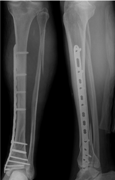
Figure 1
Postoperative radiographs after initial osteosynthesis.
Figure 2
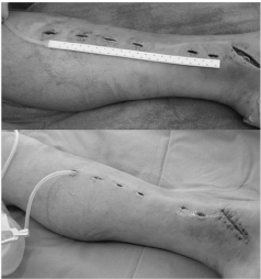
Figure 2
Top: The Jackson-Pratt drain is shown on the leg. Bottom: The drain is in place under the skin along the plate. The larger distal wound is closed.
Figure 3
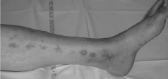
Figure 3
At seven month follow-up, there are no signs of recurrent infection.
Discussion
An acute infection following the MIPO technique for a fracture
of the distal tibia may lead the surgeon to consider opening the soft
tissues over the entire length of the plate in order to perform efficient
surgical irrigation and débridement. However, this would not only
detract from the advantages of the technique in terms of minimizing
soft tissue trauma and preserving extra-osseous blood supply to the
tibia, but also leave the plate and screws exposed and thus create
potential problems with later closure due to skin retraction, especially
in this location.
There are many benefits with the technique described in this
report. Irrigation of the infected wounds and placement of the
Jackson-Pratt drain are made through the previous MIPO incisions,
without further dissection, thus preserving the soft tissues and the
local vascular supply. The drain is made of hypo-allergenic silicone
and is soft and slippery enough to be easily pulled in and out of the
length of the wound without injuring the soft tissues and with little
pain. The design of the drain with multiple holes allows a good flow
of irrigating solution, and if correctly placed it covers the plate. It has
been shown that gently but regularly irrigating a wound with saline
is the most efficient way to reduce bacterial levels [18], and doing
so daily through the drain reduces the need for repeated visits to
the operating room for irrigation and débridement, thus reducing
hospital costs. If the patient agrees then even the definitive closure of
the wound can be done at the bedside.
Would the evolution be not optimal, the technique doesn’t
prevent a conversion to a classical treatment consisting of aggressive
débridement, lavage and hardware exchange if deemed necessary.
Technical problems can arise with use of the drain. It can become
blocked, and for this reason we recommend rinsing the wound with
at least 200 millilitres of saline each day or even more often if possible.
Although the drain could theoretically break when pulled out [19-20],
the lost part of the drain should be easily retrieved in its subcutaneous
location.
In conclusion, the use of a Jackson-Pratt drain in the setting of
acute infection following MIPO of the distal tibia is a simple and
inexpensive technique that can greatly improve the management of
this complication in the least invasive way possible.
References
- Borrelli J, Prickett W, Song E, Becker D, Ricci W. Extraosseous blood supply of the tibia and the effects of different plating techniques: a human cadaveric study. J Orthop Trauma. 2002; 16: 691-695.
- Farouk O, Krettek C, Miclau T, Schandelmaier P, Guy P, Tscherne H. Minimally invasive plate osteosynthesis and vascularity: preliminary results of a cadaver injection study. Injury. 1997; 28: A7-A12.
- Borg T, Larsson S, Lindsjo U. Percutaneous plating of distal tibial fractures. Preliminary results in 21 patients. Injury. 2004; 35: 608-614.
- Collinge C, Kuper M, Larson K, Protzman R. Minimally invasive plating of high-energy metaphyseal distal tibia fractures. J Orthop Trauma. 2007; 21: 355-361.
- Collinge C, Sanders R, DiPasquale T. Treatment of complex tibial periarticular fractures using percutaneous techniques. Clin Orthop Relat Res. 2000; 375: 69-77.
- Hasenboehler E, Rikli D, Babst R. Locking compression plate with minimally invasive plate osteosynthesis in diaphyseal and distal tibial fracture: a retrospective study of 32 patients. Injury. 2007; 38: 365-370.
- Hazarika S, Chakravarthy J, Cooper J. Minimally invasive locking plate osteosynthesis for fractures of the distal tibia--results in 20 patients. Injury. 2006; 37: 877-887.
- Helfet DL, Shonnard PY, Levine D, Borrelli J Jr. Minimally invasive plate osteosynthesis of distal fractures of the tibia. Injury. 1997; 28: A42-A47.
- Lau TW, Leung F, Chan CF, Chow SP. Wound complication of minimally invasive plate osteosynthesis in distal tibia fractures. Int Orthop. 2008; 32: 697-703.
- Maffulli N, Toms AD, McMurtie A, Oliva F. Percutaneous plating of distal tibial fractures. Int Orthop. 2004; 28: 159-162.
- Oh CW, Kyung HS, Park IH, Kim PT, Ihn JC. Distal tibia metaphyseal fractures treated by percutaneous plate osteosynthesis. Clin Orthop Relat Res. 2003; 408: 286-291.
- Pai V, Coulter G, Vishal Pai. Minimally invasive plate fixation of the tibia. Int Orthop 2007; 31: 491-496.
- Redfern DJ, Syed SU, Davies SJ. Fractures of the distal tibia: minimally invasive plate osteosynthesis. Injury. 2004; 35: 615-620.
- Vallier HA, Le TT, Bedi A. Radiographic and clinical comparisons of distal tibia shaft fractures (4 to 11 cm proximal to the plafond): plating versus intramedullary nailing. J Orthop Trauma. 2008; 22: 307-311.
- Anglen J, Watson J. Musculoskeletal infection associated with skeletal trauma. In: Stannard JP, Schmidt AH, Kregor PJ. Surgical Treatment of Orthopaedic Trauma. Thieme. 2007: 27.
- Gustillo R. Management of Infected Fractures. In: McCollister Evarts C. Surgery of the Musculoskeletal System. W.B. Saunders Company. 1990: 4432.
- Moholkar K, Ziran B. Acute Postoperative Infection. In: Bucholz RW, Heckman JD, Court-Brown C. Rockwood & Green's Fractures in Adults. Lippincott Williams & Wilkins. 2006: 580-581.
- Owens BD, White DW, Wenke JC. Comparison of irrigation solutions and devices in a contaminated musculoskeletal wound survival model. J Bone Joint Surg Am. 2009; 91: 92-98.
- Hak DJ. Retained broken wound drains: a preventable complication. J Orthop Trauma. 2000; 14: 212-213.
- Hartanto VH, Han K, Ankem M, Diamond SM. Endoscopic retrieval of retained Jackson-Pratt drain. Urology. 2001; 57: 973-974.