Case Report
A Very Rare Case of Fibrous Dysplasia Located in Odontoid Process
Hao Liu1*, Yi Yang1, Litai Ma1 and Ying Hong2
1Department of Orthopedics, Sichuan University, China
2Operation Room, Sichuan University, China
*Corresponding author: Hao Liu, Department of Orthopedics, West China Hospital, Sichuan University, Guoxuexiang, No. 37, Chengdu 610041, Sichuan Province, P. R. China
Published: 12 Sep, 2016
Cite this article as: Hao Liu. A Very Rare Case of Fibrous Dysplasia Located in Odontoid Process. Ann Clin Case Rep. 2016; 1: 1125.
Keywords: Fibrous dysplasia; Odontoid process; Cervical spine
Clinical Image
A 47-year-old female patient was admitted to our institution with the chief complaint of persistent neck pain for more than 3 months. She had no symptoms of numbness, weakness, or pain in her extremities. Noobvious abnormality was detected through neurological examination. Cervical X-rays, computed tomography (CT) scan and magnetic resonance imaging (MRI) were performed. MRI revealed a lesion in the middle part of odontoid process without involvement of the spinal cord (Figure1). Laboratory findings were within normal limits. Surgeons and radiologists all tend to the diagnosis of tumor after a heated discussion in our department. In order to confirm the character of the lesion, a biopsy surgery through anterior approach was performed. To our surprise, pathological tissue hematoxylin and eosin (HE) staining supported a diagnosis of fibrous dysplasia (Figure 2). Considering the patient had no obvious neurological abnormality, the cervical stability was not affected and the character of benign tumor, the patient was treated with conservative method with regular follow-up. Fibrous dysplasia, firstly reported by Lichtenstein in 1938, is a bone formation disorder characterized by the replacement of bone and marrow with poorly organized spicules of immature bone in a fibrous connective tissue [1]. Fibrous dysplasia can be divided into two kinds of subtypes: monostotic fibrous dysplasia and polyostotic fibrous dysplasia with or without endocrinopathy. During the past two decades, less than 40 cases of spinal fibrous dysplasia with a limited follow-up duration have been reported according to a review published in 2013 [2]. Fibrous dysplasia involving the cervical spine is rare [3]. To the best of our knowledge, this is the second report of fibrous dysplasia located in the odontoid process since Stompro et al. [4] firstly reported a case in 1989.
Figure 1
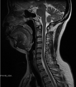
Figure 1
Magnetic resonance imaging (MRI) revealed a lesion in the middle part of odontoid process without
involvement of the spinal cord.
Figure 2
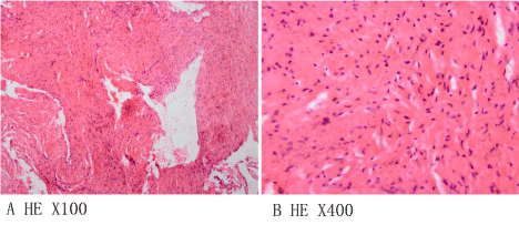
Figure 2
Pathological tissue hematoxylin and eosin (HE) staining supported
a diagnosis of fibrous dysplasia.
Acknowledgement
The patient provided informed consent for the publication of her clinical, pathological and radiological data.
References
- Bianco P, Riminucci M, Majolagbe A, Kuznetsov SA, Collins MT, Mankani MH, et al. Mutations of the GNAS1 gene, stromal cell dysfunction, and osteomalacic changes in non-McCune-Albright fibrous dysplasia of bone. J Bone Miner Res. 2000; 15: 120-128.
- Wu FL, Jiang L, Liu C, Yang SM, Wei F, Dang L, et al. Fibrous dysplasia of the mobile spine: report of 8 cases and review of the literature. Spine (Phila Pa 1976). 2013; 38: 2016-2022.
- Hakim DN, Pelly T, Kulendran M, Caris JA. Benign tumours of the bone: A review. J Bone Oncol. 2015; 4: 37-41.
- Stompro BE, Alksne JF, Press GA. Diagnosis and treatment of an odontoid fracture in a patient with polyostotic fibrous dysplasia: case report. Neurosurgery 1989; 24: 905-909.